
Table 1
TaqMan assays used in this study
| Symbol | Name | Assay No. |
|---|---|---|
| CHRM2 | cholinergic receptor muscarinic 21 | Ss03383697_u1 |
| CHRM3 | cholinergic receptor muscarinic 3 | Ss03387661_u1 |
| GAPDH | glyceraldehyde-3-phosphate dehydrogenase | Ss03375435_u1 |
| ACTB | β-actin | Ss03376081_u1 |
| HPRT | hypoxanthine guanine phosphoribosyl transferase | Ss03388274_m1 |
Table 2
Distribution and intensity of M2R, M3R, and α-7 nAChR staining in the gilt uteri of the Ctrl, Sal and E. coli groups
| Receptor | M2R | M3R | α- | 7 nAChR | |||||
|---|---|---|---|---|---|---|---|---|---|
| Uterine structure | Ctrl | Sal | E. coli | Ctrl | Sal | E. coli | Ctrl | Sal | E. coli |
| Endometrium | |||||||||
| Luminal epithelium | −/+ | −/+ | −/+ | −/+/++/+++ | −/+/++/+++ | −/+/++ | −/+/++ | −/+/++ | −/+ |
| Glandular epithelium | −/+/++ | −/+/++ | −/+/++ | −/+/++/+++ | −/+/++/++ | −/+/++/+++ | −/+ | −/+ | −/+ |
| Arterial endothelium | −/+/++ | −/+/++ | −/+/++ | −/+/++ | −/+/++ | −/+/++ | −/+ | −/+ | −/+ |
| Arterial muscle layer | −/+ | −/+ | −/+ | −/+/++ | −/+/++ | −/+ | −/+ | −/+ | −/+ |
| Venous wall | −/+ | −/+ | −/+ | −/+/++ | −/+/++ | −/+/++ | −/+ | −/+ | −/+ |
| Stroma | −/+ | −/+ | −/+ | −/+ | −/+ | −/+/++ | −/+ | −/+ | −/+ |
| Myometrium | |||||||||
| Muscle circular layer | −/+/++ | −/+/++ | −/+/++ | −/+/++ | −/+/++ | −/+/++/+++ | −/+ | −/+ | −/+ |
| Muscle longitudinal layer | −/+ | −/+ | −/+/+ | −/+/++ | −/+/++ | −/+/++/+++ | −/+ | −/+ | −/+ |
| Arterial endothelium | −/+/++ | −/+/++ | −/+/++ | −/+ | −/+ | −/+ | −/+ | −/+ | −/+ |
| Arterial muscle layer | −/+ | −/+ | −/+ | −/+ | −/+ | −/+ | −/+ | −/+ | −/+ |
| Venous wall | −/+ | −/+ | −/+ | −/+ | −/+ | −/+ | −/+ | −/+ | −/+ |
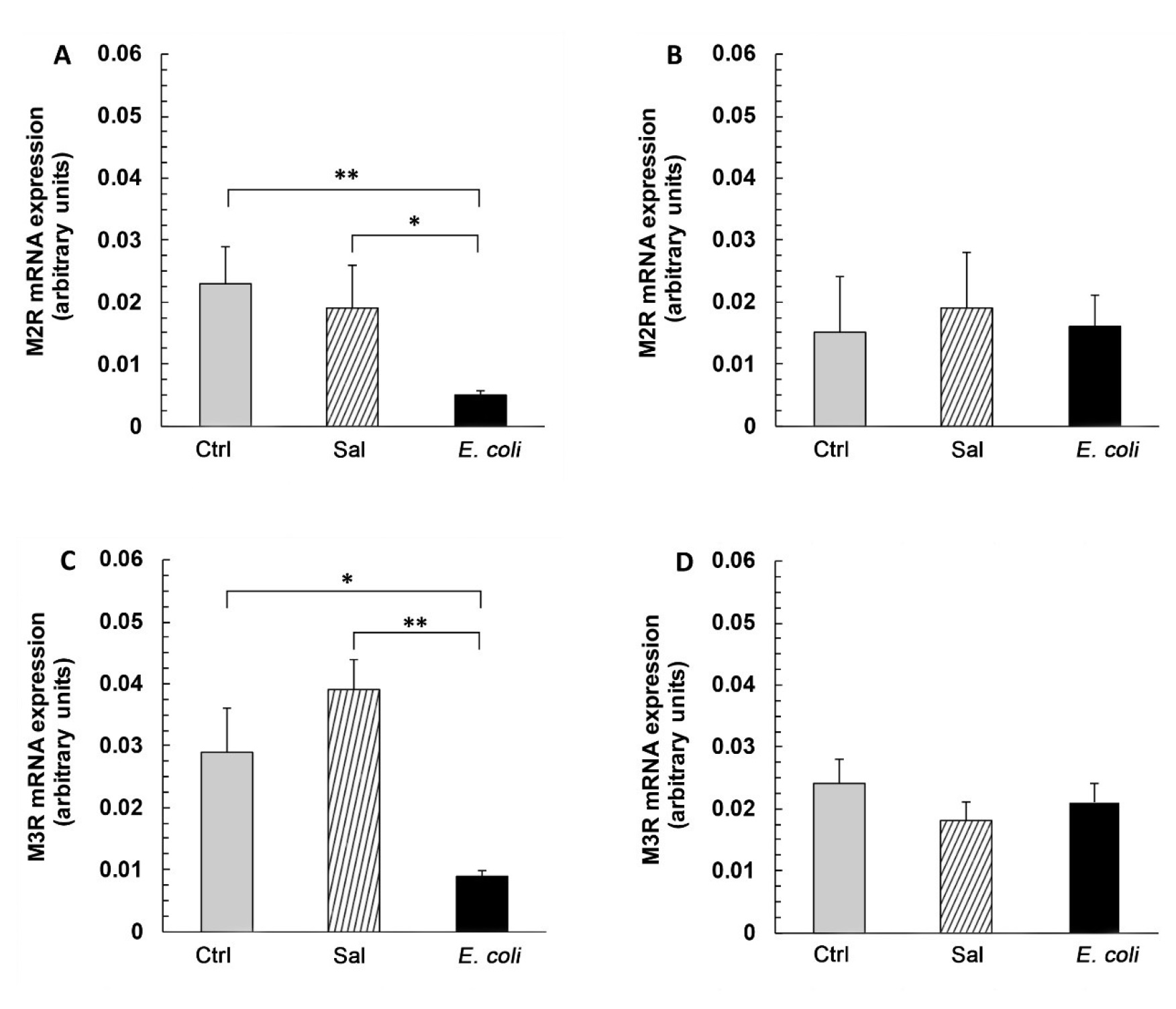
Fig. 1
Messenger RNA levels of M2R (A and B) and M3R (C and D) in the endometrium (A and C) and myometrium (B and D) of gilts from the Ctrl, Sal, and E. coli groups. Results of quantification by real-time PCR are expressed as the mean ± SEM of ratios relative to glyceraldehyde-3-phosphate dehydrogenase (GAPDH). * – P < 0.05 and ** – P < 0.01 indicate differences between groups for the same type of receptor
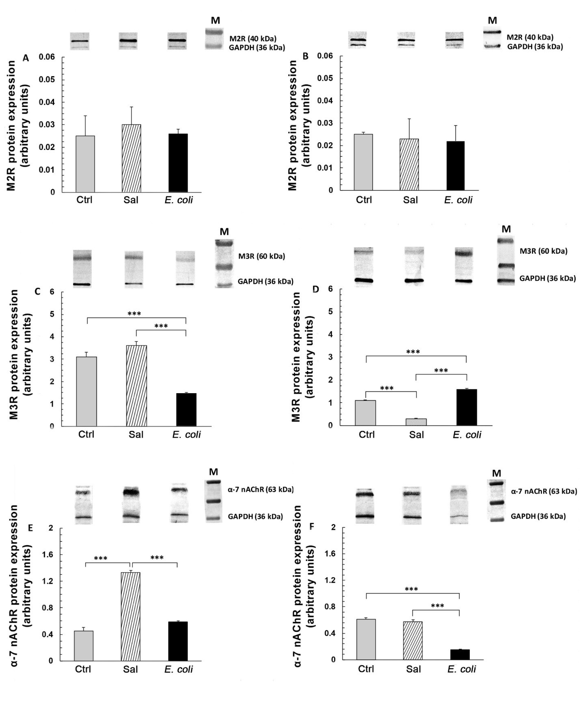
Fig. 2
Protein expression of M2R (A and B), M3R (C and D), and α-7 nAChR (E and F) in the endometrium (A, C and E) and myometrium (B, D and F) of gilts from the Ctrl, Sal and E. coli groups (n = 5 for each group). Representative blots are shown on upper panels. The results of quantification of protein contents are expressed as the mean ± SEM of ratios relative to glyceraldehyde-3-phosphate dehydrogenase (GAPDH). *** – P < 0.001 indicates differences between groups for the same type of receptor. M – marker
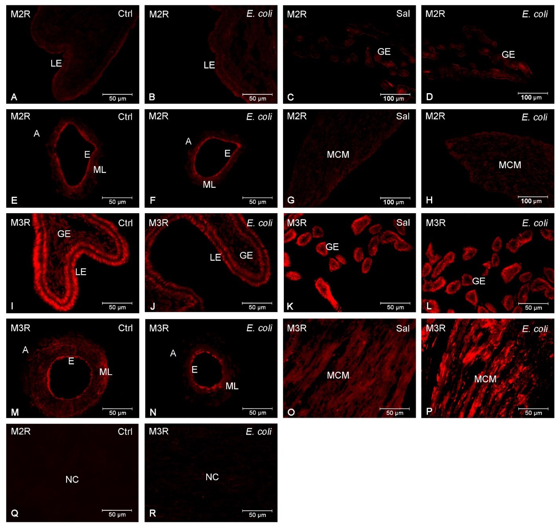
Fig. 3
Representative images of M2R (A–H) and M3R (I–P) in the uteri of gilts from the Ctrl, Sal, and E. coli groups
Image A – weak immunoexpression of M2R in the luminal epithelium of the Ctrl group
Image B – weak immunoexpression of M2R in the luminal epithelium of the E. coli group
Image C – moderate and weak immunoexpression of M2R in the glandular epithelium of the Sal group
Image D – moderate and weak immunoexpression of M2R in the glandular epithelium of the E. coli group
Image E – moderate immunoexpression of M2R in the arteries of the endometrium and in the endothelium, and weak or absent immunoexpression in the muscle layer of the Ctrl group
Image F – moderate immunoexpression of M2R in the arteries of the endometrium and in the endothelium, and weak or absent
immunoexpression in the muscle layer of the E. coli group
Image G – weak or absent immunoexpression of M2R in muscle cells of the myometrium of the Sal group
Image H – weak or absent immunoexpression of M2R in muscle cells of the myometrium of the E. coli group
Image I – strong immunoexpression of M3R in the luminal epithelium and moderate immunoexpression in the glandular epithelium of the Ctrl group
Image J – weak immunoexpression of M3R in the luminal epithelium and glandular epithelium of the E. coli group
Image K – strong immunoexpression of M3R in the glandular epithelium of the Sal group
Image L – strong immunoexpression of M3R in the glandular epithelium of the E. coli group
Image M – moderate immunoexpression of M3R in the arteries of the endometrium, endothelium, and muscle layer of the Ctrl group
Image N – weak immunoexpression of M3R in the arteries of the endometrium, endothelium, and muscle layer of the E. coli group
Image O – moderate and weak immunoexpression of M3R in the muscle cells of the myometrium of the Sal group
Image P – strong immunoexpression of M3R in the muscle cells of the myometrium of the E. coli group
Image Q – negative control for M2R after replacing the primary antibody with rabbit normal IgG
Image R – negative control for M3R after replacing the primary antibody with rabbit normal IgG
A – artery, E – endothelium, GE – glandular epithelium, LE – luminal epithelium, MCM – muscle cells of myometrium, ML – muscle layer, NC – negative control
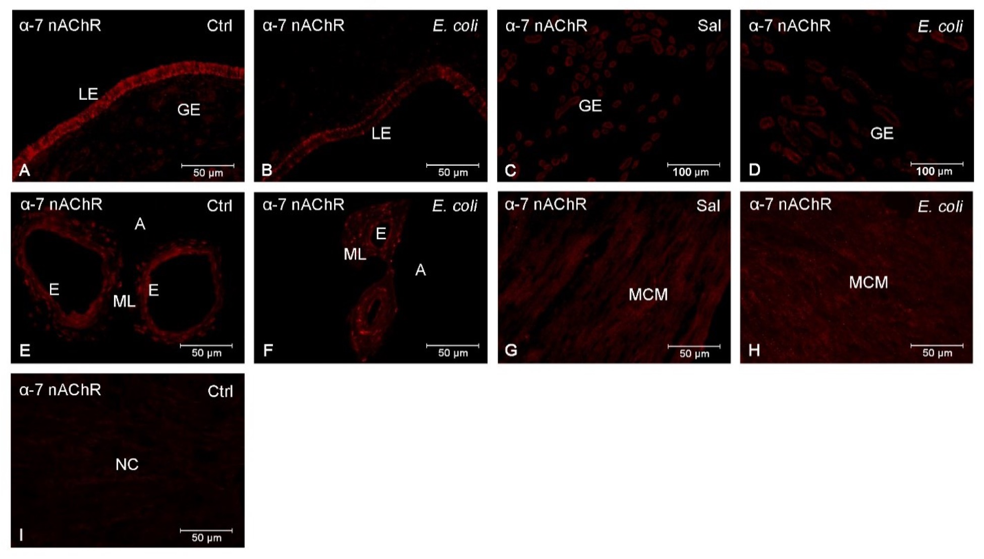
Fig.4
Representative images of α-7 nAChR in the uteri of gilts from the control (Ctrl), saline (Sal) and E. coli (E. coli) groups Image A – moderate immunoexpression of α-7 nAChR in the luminal epithelium and weak immunoexpression in the glandular epithelium of the Ctrl group
Image B – weak immunoexpression of α-7 nAChR in the luminal epithelium of the E. coli group
Image C – weak immunoexpression of α-7 nAChR in the glandular epithelium of the Sal group
Image D – weak immunoexpression of α-7 nAChR in the glandular epithelium of the E. coli group
Image E – weak or absent immunoexpression of α-7 nAChR in the arteries of the endometrium, endothelium, and muscle layer of the Ctrl group
Image F – weak or absent immunoexpression of α-7 nAChR in the arteries of the endometrium, endothelium, and muscle layer of the E. coli group
Image G – weak immunoexpression of α-7 nAChR in the muscle cells of the myometrium of the Sal group
Image H – weak immunoexpression of α-7 nAChR in the muscle cells of the myometrium of the E. coli group
Image I – negative control for α-7 nAChR after replacing the primary antibody with rabbit normal IgG
A – artery, E – endothelium, GE – glandular epithelium, LE – luminal epithelium, MCM – muscle cells of myometrium, ML – muscle layer, NC – negative control