
Table 1
Experimental and control group animal basic information
| No. | Variety | Age (years) | Gender | Spaying status | Tumour type | Disease stage |
|---|---|---|---|---|---|---|
| T-1 | Chinese pastoral cat | 10 | Female | Unspayed | Mammary gland carcinoma | Without metastasis |
| T-2 | Chinese pastoral cat | 10 | Female | Unspayed | Mammary gland carcinoma | Without metastasis |
| T-3 | Chinese pastoral cat | 9 | Female | Unspayed | Mammary gland carcinoma | Without metastasis |
| T-4 | Chinese pastoral cat | 9 | Female | Unspayed | Mammary gland carcinoma | Without metastasis |
| T-5 | Chinese pastoral cat | 8 | Female | Unspayed | Mammary gland carcinoma | Without metastasis |
| T-6 | Chinese pastoral cat | 7 | Female | Unspayed | Mammary gland carcinoma | Without metastasis |
| C-1 | Chinese pastoral cat | 10 | Female | Unspayed | Healthy control | / |
| C-2 | Chinese pastoral cat | 9 | Female | Unspayed | Healthy control | / |
| C-3 | Chinese pastoral cat | 9 | Female | Unspayed | Healthy control | / |
| C-4 | Chinese pastoral cat | 8 | Female | Unspayed | Healthy control | / |
| C-5 | Chinese pastoral cat | 8 | Female | Unspayed | Healthy control | / |
| C-6 | Chinese pastoral cat | 8 | Female | Unspayed | Healthy control | / |
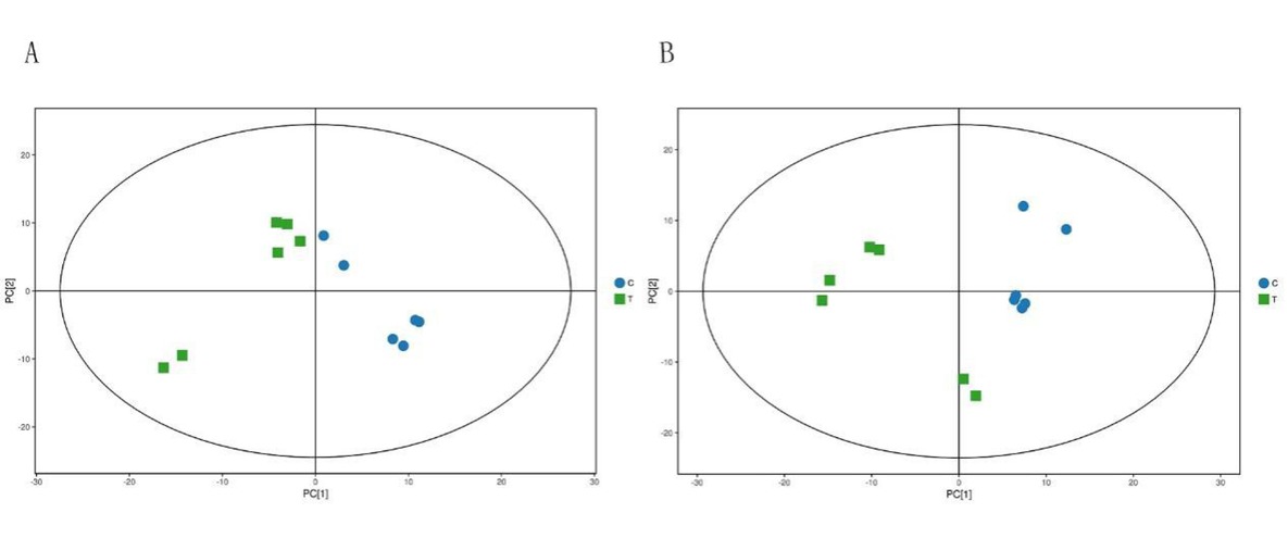
Fig. 1
PCA score diagram in NEG mode and POS mode
In the figure, the ellipse represents the 95% confidence interval, and the abscissa PC [1] and ordinate PC [2] represent the score of the principal component ranking first and second, respectively. The scatter shapes and colours represent the different experimental groups
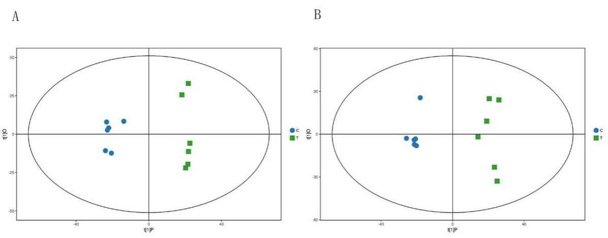
Fig. 2
OPLS-DA score in the NEG mode and POS mode
In the figure, the ellipse represents the 95% confidence interval, and the abscissa t[1]P represents the predicted principal component score of the first principal component. The ordinate t[1]O represents the orthogonal principal component score, and the scatter shapes and colours represent the different experimental groups
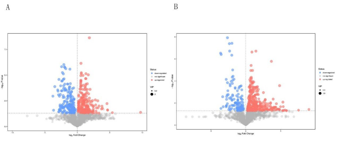
Fig. 3
Volcano map of NEG (A) and POS (B) modes differential metabolite screening
The abscissa represents the fold-change of the group comparing each substance (taking the logarithm base 2). The ordinate represents the P-value of the Student’s t-test (taking the negative logarithm base 10)
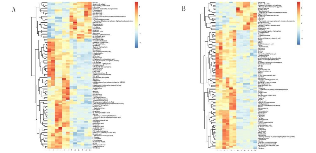
Fig. 4
HCA analysis results in NEG (A) and POS (B) modes
Table 2
Differential metabolites in feline mammary carcinomas based on LC-MS
| No. | Substance name | VIP | P | Variation trend | Detection mode |
|---|---|---|---|---|---|
| 1 | L-glutamate | 1.906599493 | 9.39957E−06 | ↑ | NEG |
| 2 | L-alanine | 1.870339069 | 9.66197E−05 | ↑ | NEG |
| 3 | Glycerol 3-phosphate | 1.813604415 | 0.001128205 | ↑ | NEG |
| 4 | Succinate | 1.790389164 | 0.000554729 | ↑ | NEG |
| 5 | 20-hydroxy-PGE2 | 1.786772601 | 2.17846E−05 | ↑ | NEG |
| 6 | Fosfomycin | 1.779219395 | 0.002655475 | ↑ | NEG |
| 7 | 3-methyluridine | 1.764311315 | 0.000277439 | ↑ | NEG |
| 8 | N-acetyl-L-alanine | 1.720196947 | 0.000240845 | ↑ | NEG |
| 9 | Choline | 1.877285980 | 2.37754E−07 | ↑ | POS |
| 10 | Trigonelline | 1.822938835 | 0.001548866 | ↑ | POS |
| 11 | Ile-asn | 1.806880095 | 1.10469E−05 | ↑ | POS |
| 12 | Arachidonic acid (peroxide free) | 1.772611472 | 0.001126785 | ↑ | POS |
| 13 | S-methyl-5′-thioadenosine | 1.770146402 | 0.000360333 | ↑ | POS |
| 14 | Creatinine | 1.751825211 | 5.23436E−05 | ↑ | POS |
| 15 | L-histidinol | 1.74002669 | 0.000841015 | ↑ | POS |
| 16 | Guanidine acetic acid | 1.698257167 | 0.000546455 | ↑ | POS |
| 17 | Cytosine | 1.662976359 | 0.000112223 | ↑ | POS |
| 18 | Inosine | 1.481481111 | 0.002446273 | ↓ | NEG |
| 19 | Adenine | 1.792517828 | 0.001575227 | ↑ | NEG |
| 20 | Hypoxanthine | 1.730804616 | 0.00067303 | ↑ | POS |
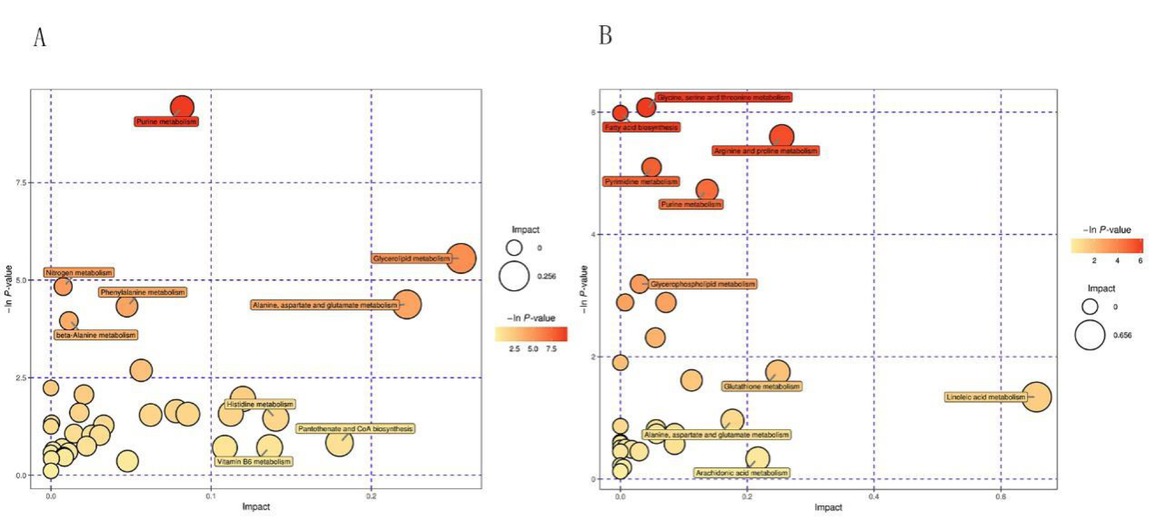
Fig. 5
Bubble diagram of the metabolic pathway analysis in NEG and POS mode
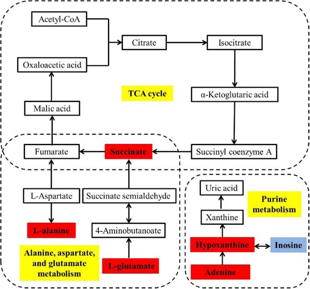
Fig. 6
Interaction network diagram of six biomarker differential metabolites. Red indicates that the metabolites are up regulated in group T, blue indicates that the metabolite is down regulated in group T, yellow indicates metabolic pathways, and metabolites without a coloured background are the upstream and downstream substances that are differentially metabolised
Table 3
MRM target metabolomic validation
| Substance name | LC-MS non-target metabolomics results | MRM target metabolomics results | ||||
|---|---|---|---|---|---|---|
| Fold change | VIP | P | Variation trend (T C) | T group final concentration (average) | C group final concentration (average) | |
| S-methyl-5′-thioadesine | 2.879152807 | 1.770146402 | 0.000360333 | ↑ | 0.011 | 0.003 |
| Creatinine | 1.632513451 | 1.751825211 | 5.23436E−05 | ↑ | 42.117 | 10.13 |
| Cytosine | 1.981265029 | 1.662976359 | 0.000112223 | ↑ | 1.383 | 0.49 |
| Choline | 1.836734369 | 1.877285980 | 2.37754E−07 | ↑ | 4.381 | 0.71 |
| Betaine nicotinate | ||||||
| (Trigonelline) | 3.178675095 | 1.822938835 | 0.001548866 | ↑ | 1.915 | 0.32 |
| L-alanine | 1.891848591 | 1.870339069 | 9.66197E−05 | ↑ | 190.229 | 94.83 |
| L-glutamic acid | 5.129593730 | 1.533880516 | 0.019410600 | ↑ | 30.959 | 3.65 |