
Table 1
Species distribution of fresh dropping samples and PCR results
| Companion birds (n) | Age (months) | Number of samples by bird seller | ||||||||||||
|---|---|---|---|---|---|---|---|---|---|---|---|---|---|---|
| A* | B | C | D | E | F | G | H | I | J | K | L | M | ||
| Agapornis sp. (3) | 60 | 3 | ||||||||||||
| A. ararauna (1) | 48 | 1 | ||||||||||||
| M. undulatus (106) | 2–12 | 11 | 5 | 5 | 10 | 2 | 7 | 10 | 15 | 10 | 8 | 8 | 7 | 8 |
| N. hollandicus (2) | 24 | 2 | ||||||||||||
| P. erithacus (1) | 36 | 1 | ||||||||||||
| Total (113) | 12 | 5 | 5 | 14 | 2 | 9 | 10 | 15 | 10 | 8 | 8 | 7 | 8 | |
Table 2
Primers used in the study, target region, and amplicon lengths
| Primer names | Sequence (5′–3′) | Target region | Size (bp) | References |
|---|---|---|---|---|
| BFDV-seq-F | TTAACAACCCTACAGACGGCGA | replication associated | ||
| BFDV-seq-R | GGCGGAGCATCTCGCAATAAG | protein (rep) gene | 605 | (21) |
| APV-Ot-2,105-F | CAGCACAGAGGTACCGTGTT | VP1 gene | 831 | (1) |
| APV-Ot-2,846-R | ATCAGAGCCCTGCATGCTTT |
Table 3
Species distribution of dropping swab samples positive for APV, PBFDV, and APV & BFDV by PCR
| Companion bird | Only APV Positive/total examined (%) | Only BFDV Positive/total examined (%) | APV & BFDV Positive/total examined (%) |
|---|---|---|---|
| Agapornis sp. | 0/3 (0) | 0/3 (0) | 0/3 (0) |
| A. ararauna | 0/1 (0) | 0/1 (0) | 0/1 (0) |
| M. undulatus | 40/106 (37.7) | 11/106 (10.4) | 14/106 (13.2) |
| N. hollandicus | 1/2 (50) | 1/2 (50) | 0/2 (0) |
| P. erithacus | 0/1 (0) | 0/1 (0) | 0/1 (0) |
| Total | 41/113 (36.3) | 12/113 (10.6) | 14/113 (12.4) |
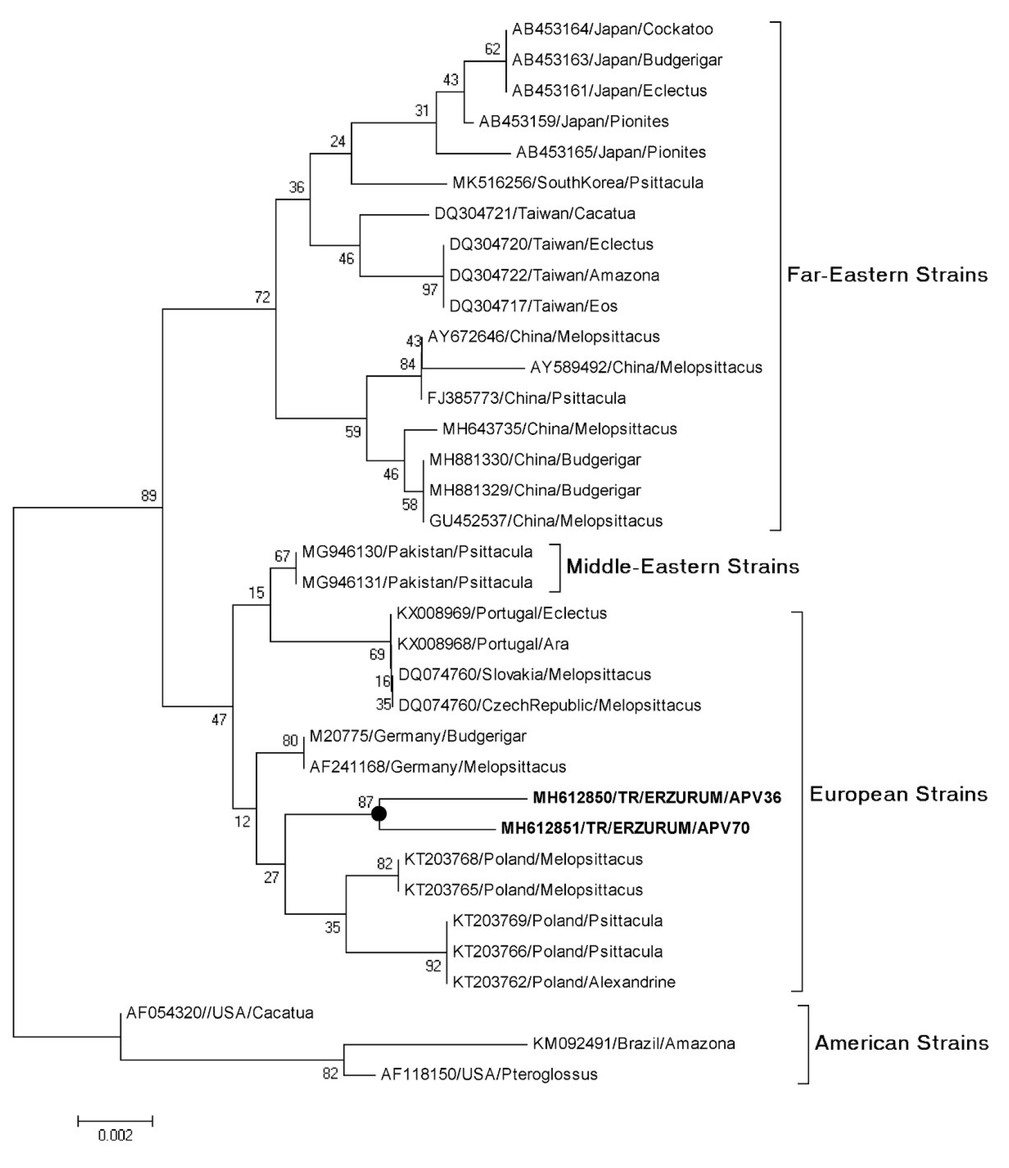
Fig. 1
Phylogenetic tree of different avian Polyomavirus (APV) strains generated using the maximum likelihood method in MEGA v X. The percentage of replicate trees in which the associated taxa clustered together in the bootstrap test (1,000 replicates) is shown next to the branches. The evolutionary distances were computed using the maximum likelihood method and are in the units of base substitutions per site. Codon positions included were 1st + 2nd + 3rd + noncoding. The analysis involved 33 nucleotide sequences
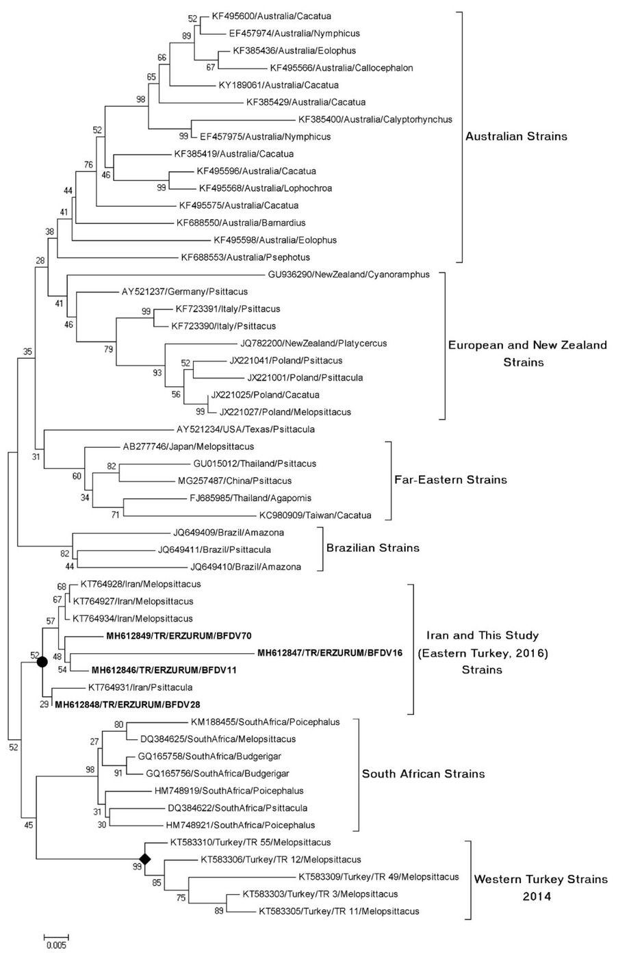
Fig. 2
Phylogenetic tree of different Circovirus strains generated using the maximum likelihood methods in MEGA v X. The percentage of replicate trees in which the associated taxa clustered together in the bootstrap test (1,000 replicates) are shown next to the branches. The evolutionary distances were computed using the maximum composite likelihood method and are in the units of base substitutions per site. The tree is drawn to scale, with branch lengths measured in the number of substitutions per site. The analysis involved 49 nucleotide sequences. Codon positions included were 1st + 2nd + 3rd + noncoding