
Table 1
Primer sequences used for qRT-PCR
| NCBI reference sequence | Gene | Forward primer (5´→3´) | Reverse primer (5´→3´) |
|---|---|---|---|
| NM_001003142.2 | GAPDH | AGTCAAGGCTGAGAACGGGAAA | TCCACAACATACTCAGCACCAGC |
| NM_001002949.1 | BCL2 | CATGCCAAGAGGGAAACACCAGAA | GTGCTTTGCATTCTTGGATGAGGG |
| NM_001003011.1 | BAX | TTCCGAGTGGCAGCTGAGATGTTT | TGCTGGCAAAGTAGAAGAGGGCAA |
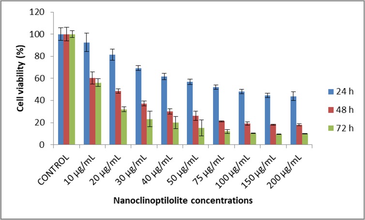
Fig. 1
Effect of nanoclinoptilolite on cell viability of canine OSA D-17
Table 2
Effect of nanoclinoptilolite on cell viability of canine D-17 OSA. The results represent the mean ± standard deviation
| Nanoclinoptilolite concentration (μg/mL) | 24 h | 48 h | 72 h | ||||||
|---|---|---|---|---|---|---|---|---|---|
| % Viability | X | P | % Viability | X | P | % Viability | X | P | |
| Control | 100 ± 5.66 | a | 100 ± 6.09 | a | 100 ± 3.15 | a | |||
| 10 | 92.62 ± 8.29 | a | >0.05 | 60.24 ± 5.50 | b | *** | 56.08 ± 3.57 | b | *** |
| 20 | 81.45 ± 5.11 | b | *** | 48.50 ± 1.83 | c | *** | 31.95 ± 2.24 | c | *** |
| 30 | 69.35 ± 2.24 | c | *** | 37.27 ± 2.25 | d | *** | 23.23 ± 6.89 | cd | *** |
| 40 | 61.66 ± 3.01 | cd | *** | 30.12 ± 2.23 | de | *** | 19.87 ± 5.45 | de | *** |
| 50 | 56.88 ± 2.45 | de | *** | 26.16 ± 4.14 | ef | *** | 15.14 ± 7.29 | def | *** |
| 75 | 51.92 ± 2.02 | def | *** | 21.19 ± 0.42 | fg | *** | 12.02 ± 1.70 | ef | *** |
| 100 | 48.08 ± 1.87 | ef | *** | 19.14 ± 1.55 | fg | *** | 10.31 ± 0.26 | ef | *** |
| 150 | 44.60 ± 1.80 | f | *** | 18.19 ± 0.53 | g | *** | 9.41 ± 0.15 | f | *** |
| 200 | 43.80 ± 3.91 | f | *** | 18.00 ± 0.74 | g | *** | 9.92 ± 0.28 | f | *** |
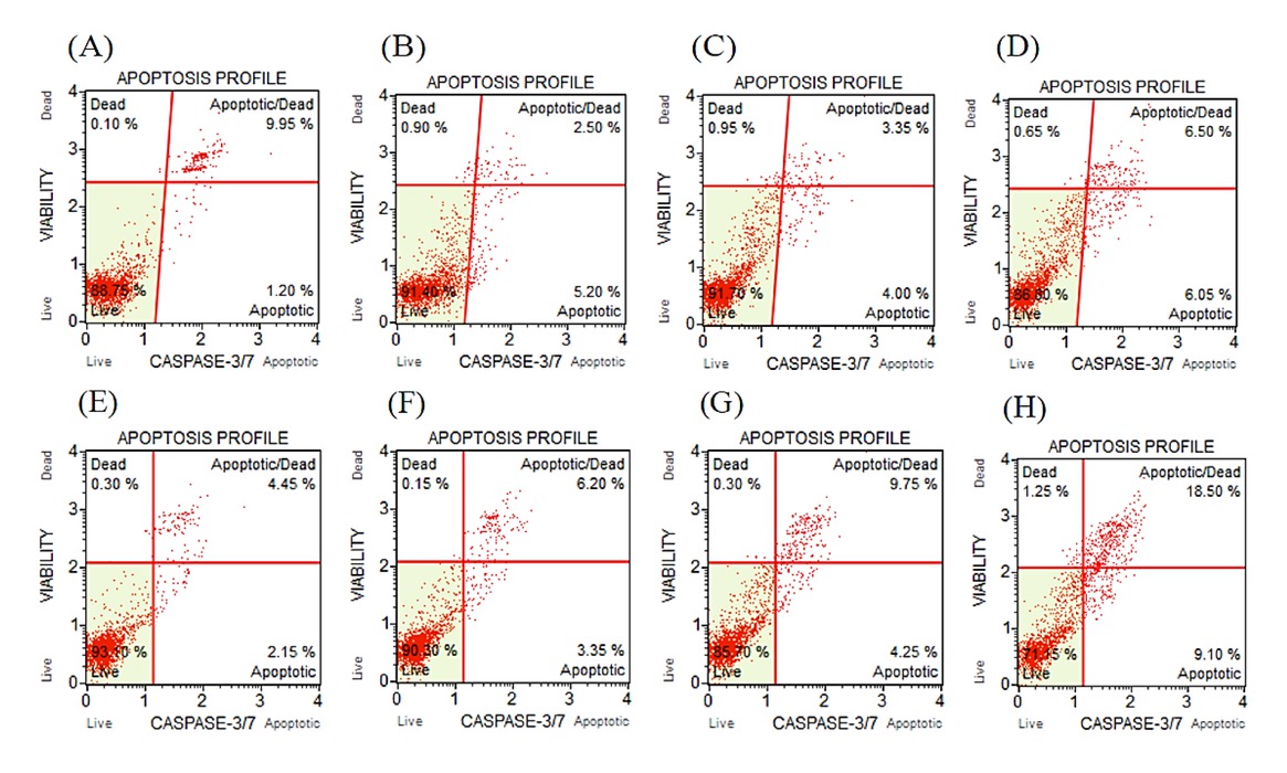
Fig. 2
Caspase-3 and -7 activity after treatment with different concentrations of nanoclinoptilolite in canine D-17 OSA cells. Treatment over 24 h: A – control; B – 10 μg/mL; C– 20 μg/mL; D – 30 μg/mL. Treatment over 48 h: E – control; F – 10 μg/mL; G – 20 μg/mL; H– 30 μg/mL
Table 3
Live/apoptotic/necrotic cell ratios in canine D-17 OSA cells treated for 24 h with 10, 20, and 30 μg/mL of nanoclinoptilolite and in control group cells
Table 4
Live/apoptotic/necrotic cell ratios in canine D-17 OSA cells treated for 48 h with 10, 20, and 30 μg/mL of nanoclinoptilolite and in control group cells
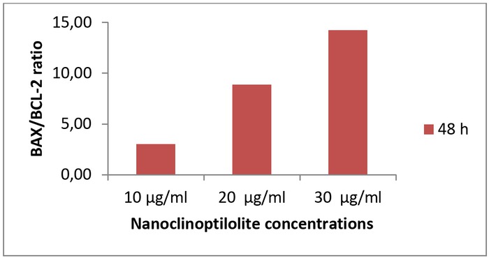
Fig. 3
Effect of nanoclinoptilolite on the BAX/BCL-2 ratio in canine D-17 OSA cells