
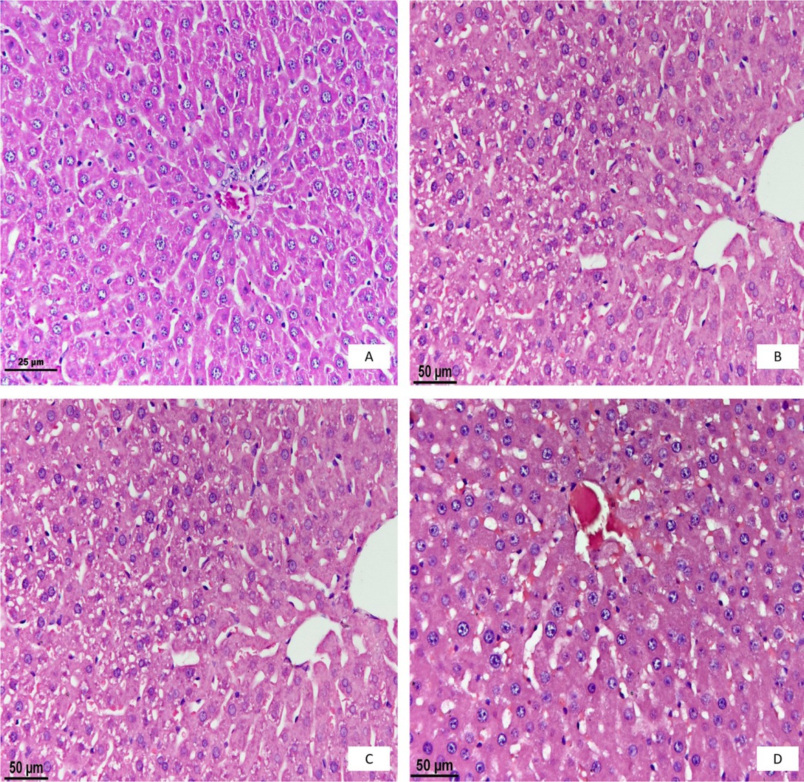
Fig. 1
Histopathological examination of the liver. A – the control group; B – the first day; C – the second day; D – the third day
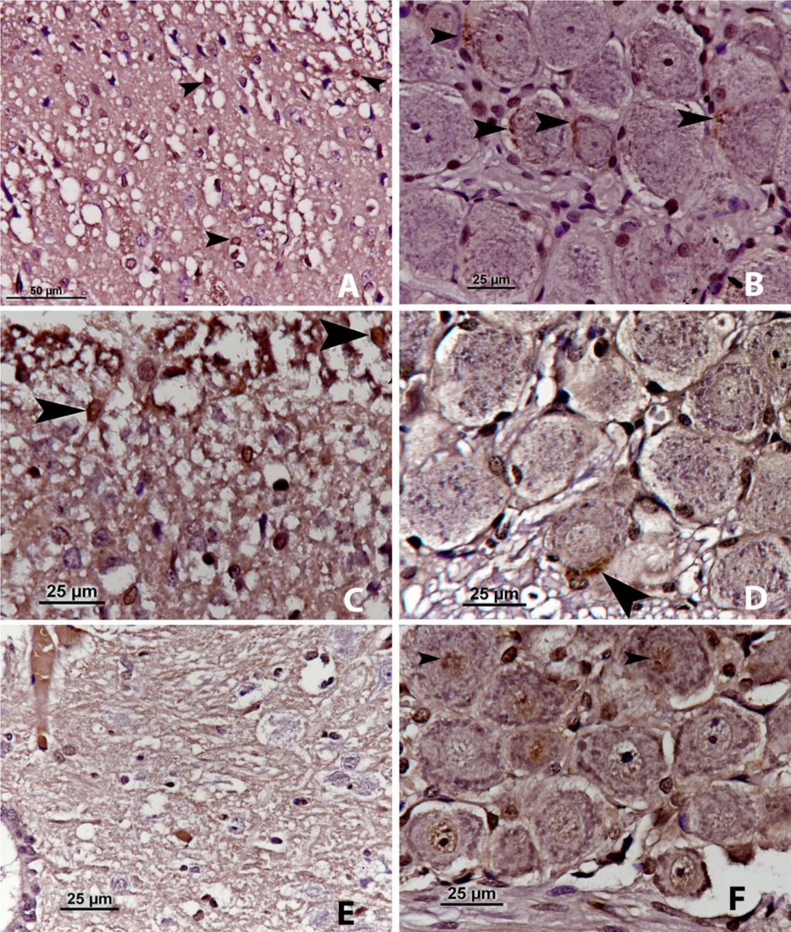
Fig. 2
nNOS immunoreactivity. A – the first day, in the spinal cord; B – the first day, in the nodose ganglion; C – the second day, in the spinal cord; D – the second day, in the nodose ganglion; E – the third day, in the spinal cord; F – the third day, in the nodose ganglion
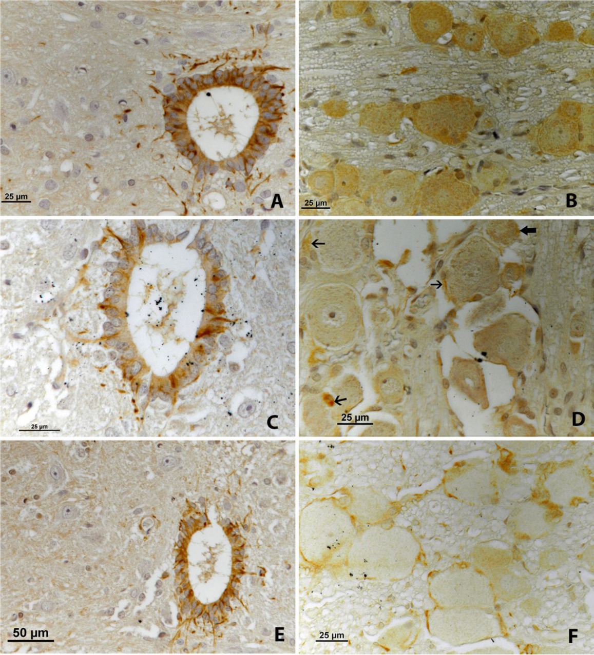
Fig. 3
iNOS immunoreactivity. A – the first day, in the spinal cord; B – the first day, in the nodose ganglion; C – the second day, in the spinal cord; D – the second day, in the nodose ganglion; E – the third day, in the spinal cord; F – the third day, in the nodose ganglion
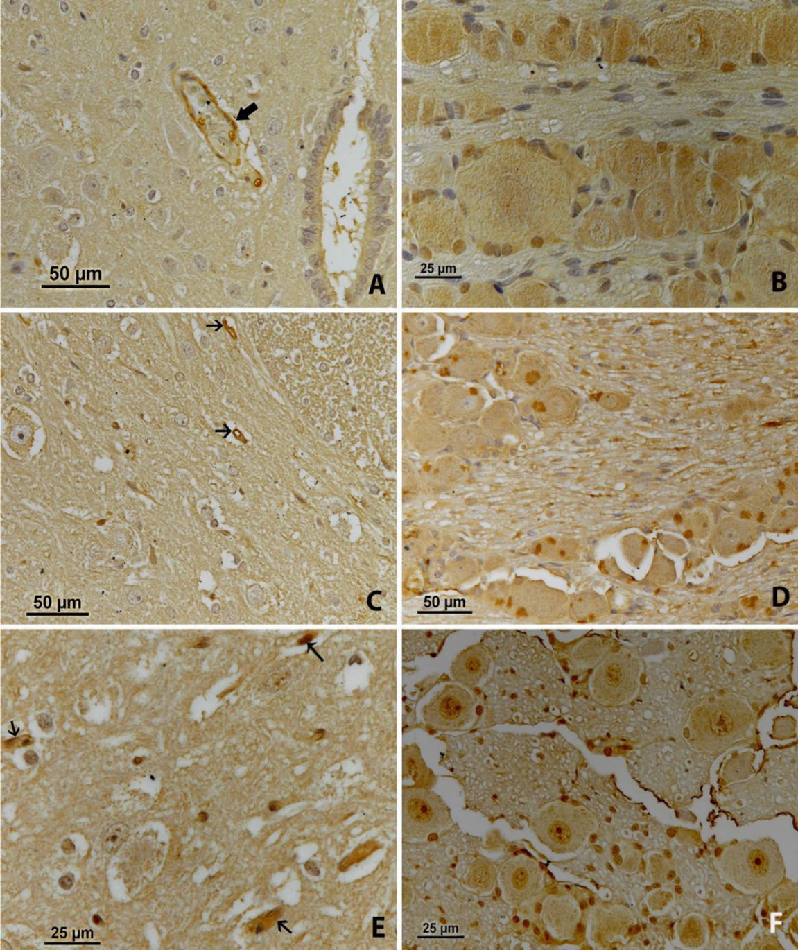
Fig. 4
eNOS immunoreactivity. A – the first day, in the spinal cord; B – the first day, in the nodose ganglion; C – the second day, in the spinal cord; D – the second day, in the nodose ganglion; E – the third day, in the spinal cord; F – the third day, in the nodose ganglion