Fig. 1
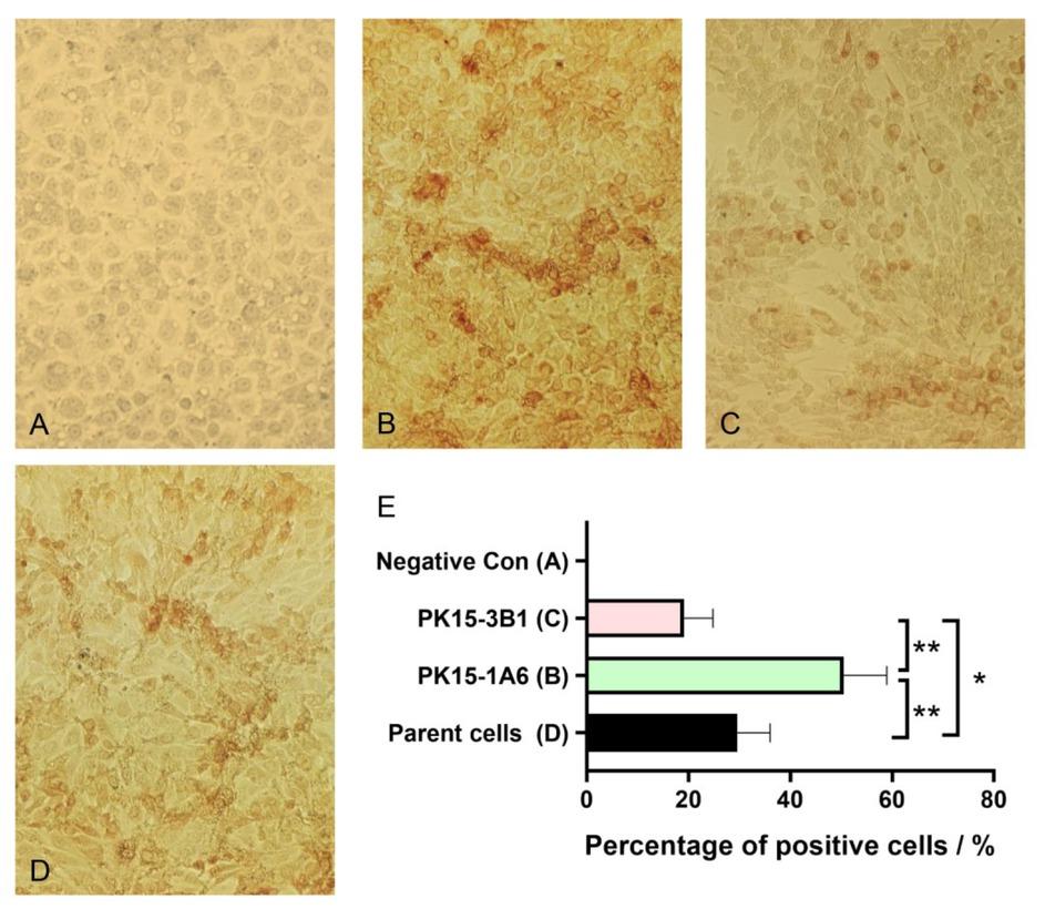
Fig. 2
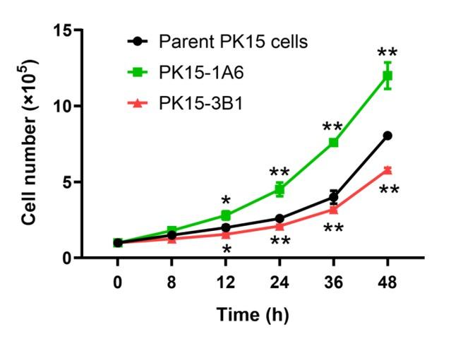
Fig. 3
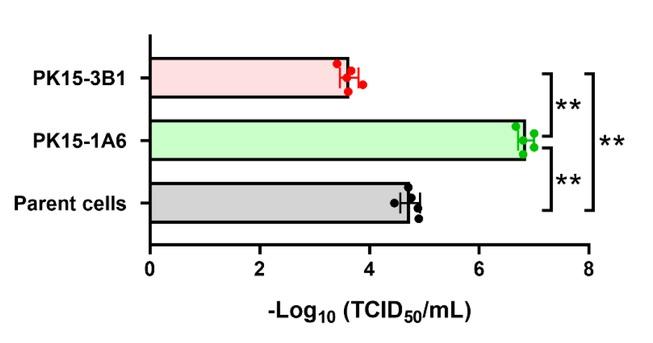
Fig. 4
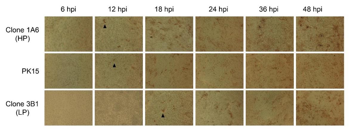
Fig. 5

© 2020 Mei Yin, Dongfang Hu, Peng Li, Lingyun Kong, Hongmei Ning, Feng Yue, Jinqing Jiang, Xuannian Wang, published by National Veterinary Research Institute in Pulawy
This work is licensed under the Creative Commons Attribution-NonCommercial-NoDerivatives 3.0 License.