
Table 1
Tumour biomarker results in healthy and OPA sheep
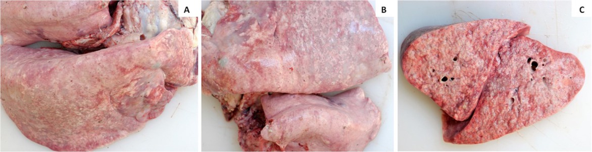
Fig. 1
Macroscopic appearance of the lungs. A – grey-white tumoural foci on a diaphragmatic lung lobe; B – large foci consisting of merged small foci; C – effusive greyish tumoural foci in cross-section of the lung
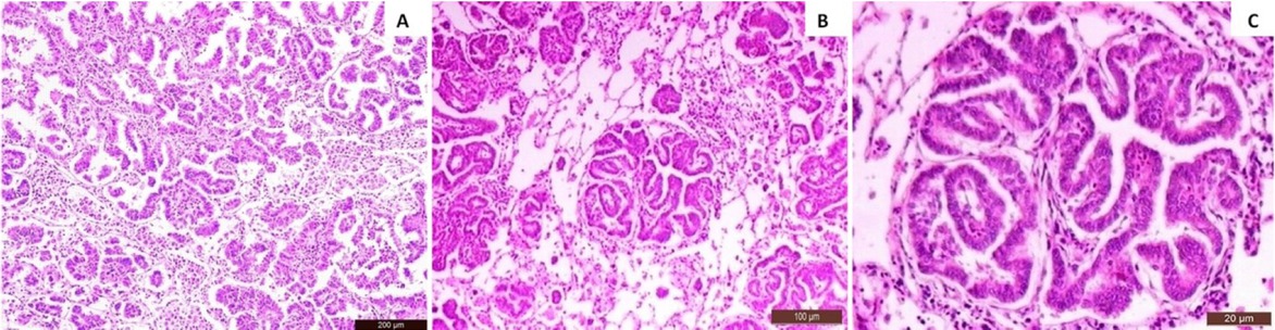
Fig. 2
Microscopic appearance of the lungs. A – type-II pneumocytes proliferating in alveolar lumen and numerous macrophages on adjacent alveoli; H&E, Bar: 200 μm; B – type-II pneumocytes proliferating in alveolar lumen and numerous macrophages on adjacent alveoli; H&E, Bar: 100 μm; C – type-II pneumocyte cells proliferating in alveolar lumens; H&E, Bar: 20 μm