
Table 1
Epidemiological variables associated with leishmaniasis in dogs from a central region of Colombia (July 2016-July 2017)
| Variables | Tolima | % | Huila | % | Total | % | OR | P < 0.05 |
|---|---|---|---|---|---|---|---|---|
| Characteristics of housing | ||||||||
| Bamboo | 1 | 33.3 | 0 | 0.0 | 1 | 9.1 | ||
| Brick | 1 | 33.3 | 4 | 50.0 | 5 | 45.5 | ||
| Wood | 0 | 0.0 | 3 | 37.5 | 3 | 27.3 | ||
| Gaudua | 1 | 33.3 | 1 | 12.5 | 2 | 18.2 | 10.5 | 0.398 |
| Sick people | 0 | 0.0 | 2 | 25.0 | 2 | 18.2 | 7 | 0.221 |
| Garbage | 1 | 33.3 | 4 | 50.0 | 5 | 45.5 | 7 | 0.221 |
| Reservoirs | 3 | 100.0 | 8 | 100.0 | 11 | 100.0 | 8.93 | 0.003 |
| House plants | 3 | 100.0 | 7 | 87.5 | 10 | 90.9 | 7 | 0.221 |
| Stream or pipes | 2 | 66.7 | 7 | 87.5 | 9 | 81.8 | 2.91 | 0.713 |
| Pests | 2 | 66.7 | 7 | 87.5 | 9 | 81.8 | 8.25 | 0.004 |
| Characteristics of canine | ||||||||
| Male | 3 | 100.0 | 7 | 87.5 | 10 | 90.9 | 7.35 | 0.007 |
| Female | 0 | 0.0 | 1 | 12.5 | 1 | 9.1 | ||
| Age | 6.3 | 3–11 | 3.6 | 2–5 | 4.4 | 2–11 | 14 | 0.173 |
| Cachexia | 2.917 | 0.713 | ||||||
| Normal | ||||||||
| Trips | 1 | 33.3 | 4 | 50.0 | 5 | 45.5 | 5.125 | 0.024 |
| Symptomatology | ||||||||
| Anamnesis | 3 | 100 | 7 | 87.5 | 10 | 90.9 | 4.95 | 0.421 |
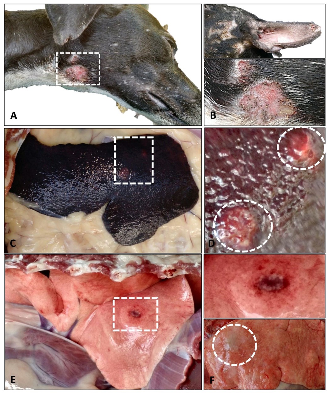
Fig. 1
Macroscopic lesions in dogs with leishmaniasis in a central region of Colombia. A, B – skin lesions (exfoliative and ulcerative dermatitis) on the head, neck, and ears (Fig. B lower panel is a magnification of the delineated square in A). C, D – splenomegaly with nodules protruding on the irregular surface of the organ (Fig. D is a magnification of the delineated square in fig. C). E, F upper panel – ulcerated nodule in the lung (Fig. F upper panel is a magnification of the delineated square in Fig. E). F lower panel – a non-ulcerated nodule protruding from the surface of the lung of a different dog
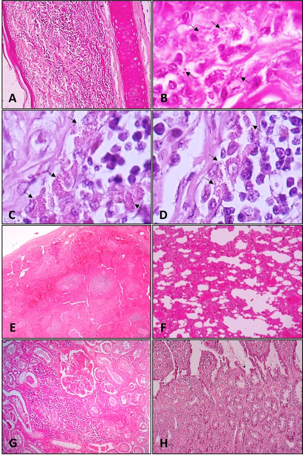
Fig. 2
Microscopic lesions in dogs with leishmaniasis in a central region of Colombia. A, B – auricular dermatitis associated with intracellular amastigotes present within macrophages (arrows). C, D – lymphadenitis showing abundant promastigotes parasites (arrows) within phagocytic mononuclear cells. E – spleen with severe depletion of lymphocytes (white pulp) and apparent hyperplasia of red pulp. F – lung with moderate to severe interstitial pneumonia. G – kidney showing interstitial nephritis with tubular degeneration. H – small intestine showing lymphoplasmacytic infiltrate
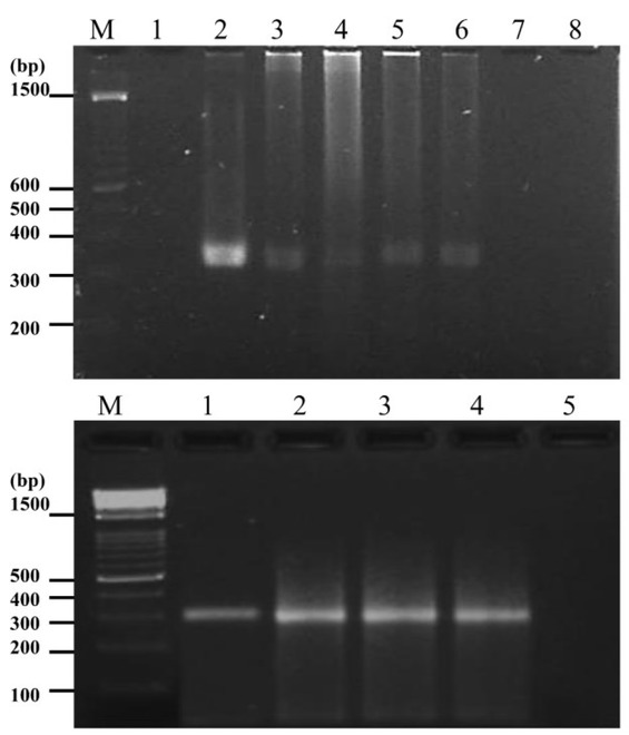
Fig. 3
PCR Amplification of a 314 bp DNA fragment of a Leishmania RNA gene. Upper panel – M:100 bp ladder. Lane 1 – negative control (without template DNA). Lane 2 – positive control of L. amazonensis. Lane 3 – spleen. Lane 4 – skin. Lanes 5, 6 – lymph nodes. Lane 7 – positive control of L. infantum. Lane 8 – positive control of L. braziliensis. Lower panel – M: 100 bp ladder. Lane 1 – positive control of L. amazonensis. Lanes 2–5 – spleen samples from three serological positive dogs