
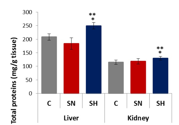
Fig. 1
Total proteins concentration in the liver and kidneys for each experimental condition (C – control; SN – sepsis normothermic; SH– sepsis hypothermic). * – different from control (P < 0.05); ** – different from SN (p < 0.05). Values are expressed as means ± SD
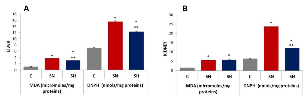
Fig. 2
GPx and SOD activity in the liver (A) and kidneys (B) for each experimental condition (C – control; SN – sepsis normothermic; SH – sepsis hypothermic). * – different from control (p < 0.05); ** – different from SN (p < 0.05). Values are expressed as means ± SD
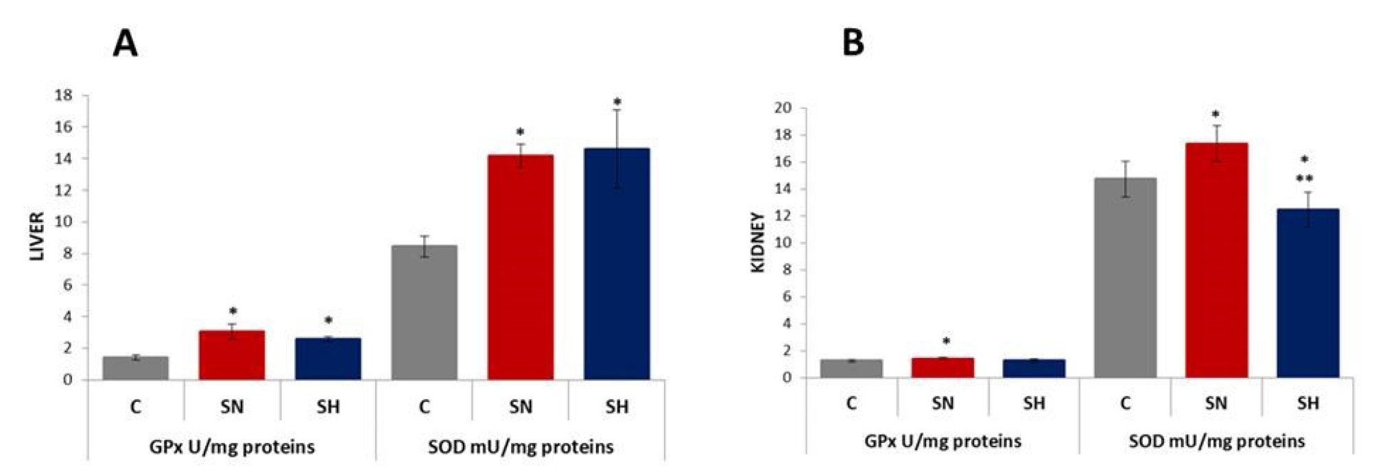
Fig. 3
MDA and DNPH concentrations in the liver (A) and kidneys (B) for each experimental condition (C – control; SN – sepsis normothermic; SH – sepsis hypothermic). * – different from control; ** – different from SN (p < 0.05). Values are expressed as means ± SD
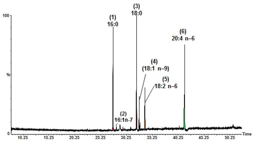
Fig. 4
GC-MS chromatogram of the FAMEs from total lipids of the liver (SH – sepsis hypothermic group) analysed with a BPx-70 column. Peaks of fatty acids: (1) palmitic (16:0); (2) palmitoleic (16:1 n–7); (3) stearic (18:0); (4) oleic (18:1 n–9); (5) linoleic (18:2 n–9); and (6) arachidonic (20:4 n–6)
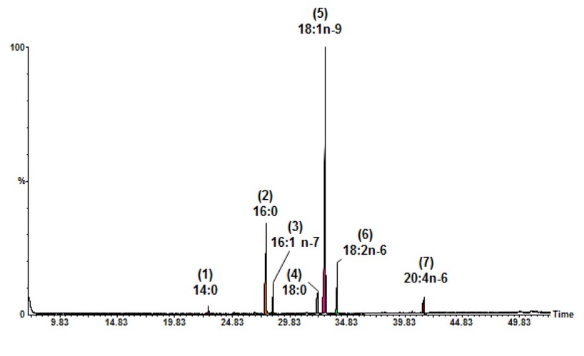
Fig. 5
GC-MS chromatogram of the FAMEs from total lipids of kidneys (SN – sepsis normothermic group) analysed with a BPx-70 column. Peaks of fatty acids: (1) myristic (14:0); (2) palmitic (16:0); (3) palmitoleic (16:1 n–7); (4) stearic (18:0); (5) oleic (18:1 n–9); (6) linoleic (18:2 n–6); and (7) arachidonic (20:4 n–6)
Table 1
Fatty acid composition of total lipids determined by GC-MS in the liver and kidneys for each experimental condition (C – control; SN – sepsis normothermic; SH – sepsis hypothermic)
| Fatty acid % a | ||||||
|---|---|---|---|---|---|---|
| Fatty acid | C | Liver SN | SH | C | Kidneys SN | SH |
| Myristic (14:0) | nd | nd | nd | 0.17 ± 0.09 | 1.01 ± 0.01 | 0.15 ± 0.01 |
| Palmitic (16:0) | 26.61 ± 1.21 | 24.48 ± 1.03 | 23.17 ± 0.98 | 7.17 ± 0.21 | 24.53 ± 1.06 | 9.71 ± 0.42 |
| Palmitoleic (16:1 n–9) | 0.16 ± 0.08 | 0.19 ± 0.06 | 0.17 ± 0.11 | 0.8 ± 0.09 | 0.21 ± 0.05 | 0.78 ± 1.01 |
| Palmitoleic (16:1 n–7) | 0.96 ± 0.12 | 0.92 ± 0.19 | 0.98 ± 0.11 | 0.26 ± 0.07 | 3.92 ± 0.24 | nd |
| Stearic (18:0) | 18.02 ± 1.13 | 29.69 ± 1.65 | 26.65 ± 1.16 | 0.53 ± 0.03 | 6.88 ± 0.29 | 0.62 ± 0.13 |
| Oleic (18:1 n–9) | 8.48 ± 0.32 | 5.15 ± 0.19 | 8.43 ± 0.28 | 69.22 ± 1.69 | 50.16 ± 1.53 | 67.62 ± 0.63 |
| Oleic (18:1 n–7) | 2.71 ± 0.13 | 1.94 ± 0.16 | 1.4 ± 0.09 | 4.01 ± 0.24 | 3.3 ± 0.52 | 3.59 ± 0.12 |
| Linoleic (18:2 n–9) | 14.12 ± 0.5 | 13.49 ± 0.29 | 12.88 ± 0.33 | 13.01 ± 0.31 | 9.85 ± 0.22 | 13.4 ± 0.52 |
| Arachidonic (20:4 n–6) | 28.94 ± 0.89 | 24.15 ± 0.91 | 26.32 ± 1.41 | 4.83 ± 0.12 | 0.14 ± 0.06 | 4.12 ± 0.16 |
[i] a – % of total fatty acids; nd – not detected. Values are mean ± standard deviation of samples analysed individually in triplicate
Table 2
Statistical analysis of fatty acid distribution in total lipids for each experimental condition (C – control; SN – sepsis normothermic; SH – sepsis hypothermic)
| Fatty acid/% a | ||||||
|---|---|---|---|---|---|---|
| Type of tissues/ | group | Σ SFAs | Σ MUFAs | Σ PUFAs | Σ UFAs | Σ UFAs / Σ SFAs |
| C | 44.63 ± 1.79 | 12.31 ± 0.46 | 43.06 ± 1.46 | 55.37 ± 2.13 | 1.24 | |
| Liver | SN | 54.17 ± 1.65* | 8.19 ± 0.29* | 37.63 ± 1.38* | 50.19 ± 1.98* | 0.84* |
| SH | 49.82 ± 1.84*,** | 10.98 ± 0.36*,** | 39.19 ± 1.65 | 45.83 ± 1.73*,** | 1.007** | |
| C | 7.87 ± 0.53 | 74.29 ± 2.93 | 17.84 ± 0.55 | 92.13 ± 3.69 | 11.70 | |
| Kidneys | SN | 32.42 ± 1.27* | 57.59 ± 1.95* | 9.99 ± 0.37* | 67.58 ± 2.24* | 2.08* |
| SH | 10.48 ± 0.67*,** | 72.00 ± 2.63** | 17.52 ± 0.51 | 89.52 ± 3.33** | 8.54** | |
[i] a – % of total fatty acids; * – different from control; ** – different from SN (P < 0.05); SFAs – saturated fatty acids; MUFAs – monounsaturated fatty acid; PUFAs – polyunsaturated fatty acids; UFAs – unsaturated fatty acids. Values are mean ± standard deviation of samples analysed individually in triplicate