
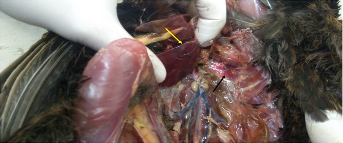
Fig. 1
Congestion, complicated by haemorrhage in the lung (black arrow) and focal hepatic necrosis (yellow arrow)
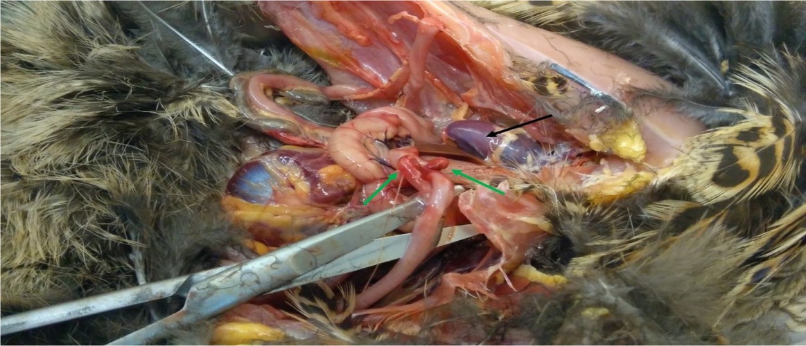
Fig. 2
Diffuse subserosal haemorrhages in the caeca (green arrows) and dystrophic and necrotic changes in the heart (black arrow)
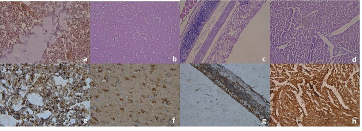
Fig. 3
Histopathological and IHC changes: a) lung – haemorrhagic pneumonia, oedema, emphysema, and atelectasis, 100 ×; b) brain – perivascular and pericellular oedema, 200 ×; c) trachea – acute tracheitis, with epithelial desquamation, 200 ×; d) myocardium – interstitial oedema, 100 ×; e) lung – IHC expression of anti-influenza A virus nucleoprotein antibody, 200 ×; f) brain – IHC expression of anti-influenza A virus nucleoprotein antibody, 200 ×; g) trachea – IHC expression of anti-influenza A virus nucleoprotein antibody, 100 ×; h) myocardium – IHC expression of anti-influenza A virus nucleoprotein antibody, 200 ×