
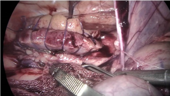
Fig. 1
The clamped artery was released two seconds after suturing to confirm haemostasis. The two haemostatic clips were released completely when there was no haemorrhage
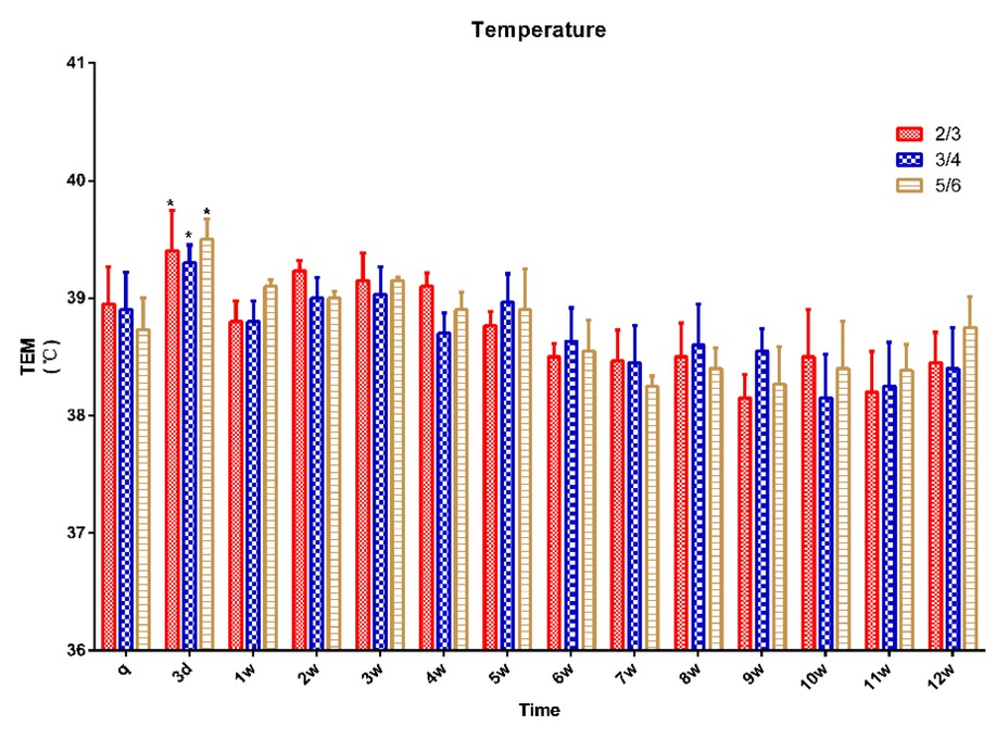
Fig. 2
Rectal temperature in 15 pigs (n = 5 for ⅔ group, n = 5 for ¾ group, n = 5 for ⅚ group) q – preoperative baseline
* p < 0.05 compared with the preoperative baseline. A p < 0.05 compared with value of ⅔ group at the same time point. B p < 0.05 compared with value of ¾ group at the same time point
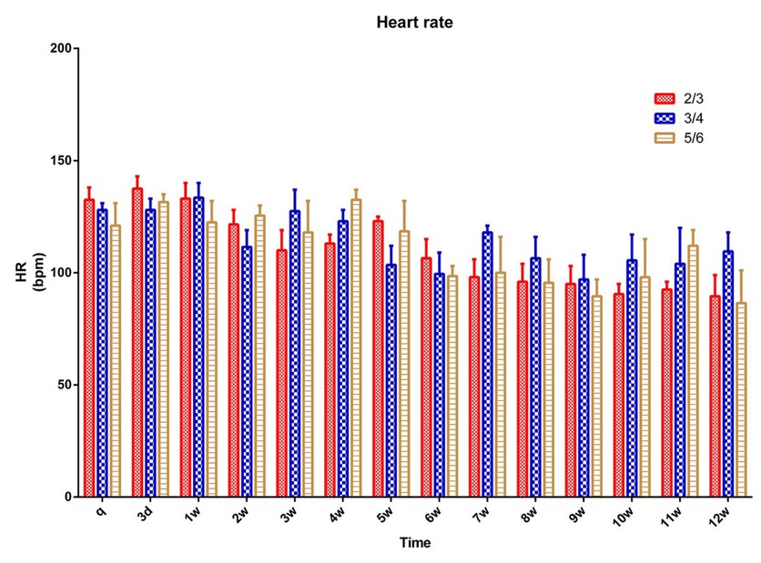
Fig. 3
Heart rate in 15 pigs (n = 5 for ⅔ group, n = 5 for ¾ group, n = 5 for ⅚ group) q – preoperative baseline
* p < 0.05 compared with the preoperative baseline. A p < 0.05 compared with value of ⅔ group at the same time point. B p < 0.05 compared with value of ¾ group at the same time point
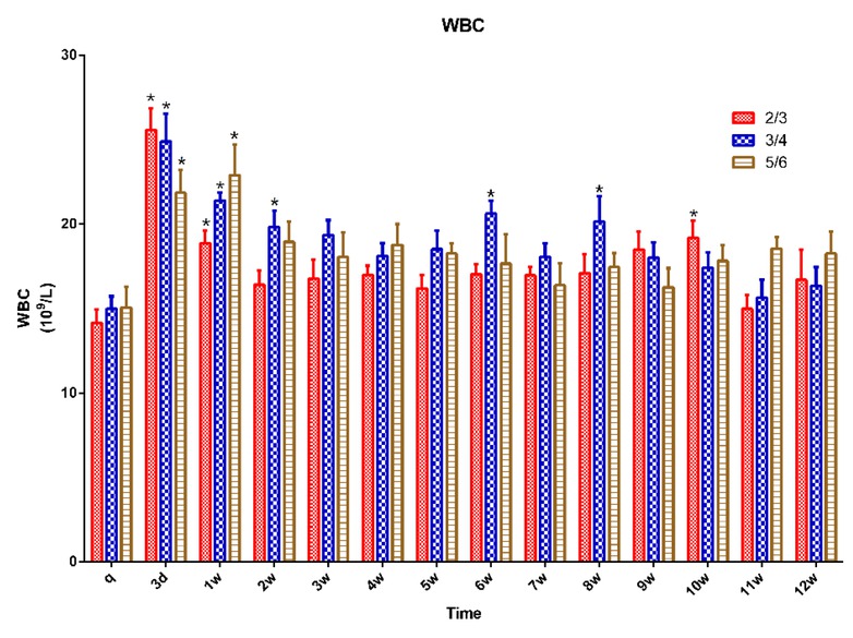
Fig. 4
WBC in 15 pigs (n = 5 for ⅔ group, n = 5 for ¾ group, n = 5 for ⅚ group) q – preoperative baseline
* p < 0.05 compared with the preoperative baseline. A p < 0.05 compared with value of ⅔ group at the same time point. B p < 0.05 compared with value of ¾ group at the same time point
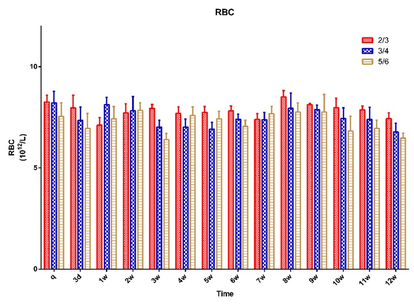
Fig. 5
RBC in 15 pigs (n = 5 for ⅔ group, n = 5 for ¾ group, n = 5 for ⅚) group q – preoperative baseline
* p < 0.05 compared with the preoperative baseline. A p < 0.05 compared with value of ⅔ group at the same time point. B p < 0.05 compared with value of ¾ group at the same time point ???
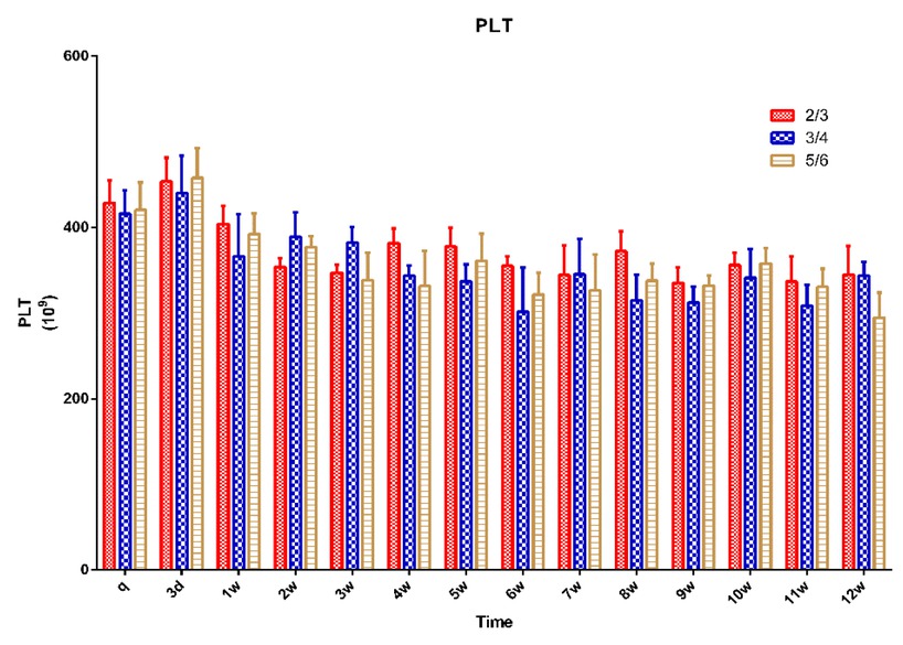
Fig. 6
PLT in 15 pigs (n = 5 for ⅔ group, n = 5 for ¾ group, n = 5 for ⅚ group) q – preoperative baseline
* p < 0.05 compared with the preoperative baseline. A p < 0.05 compared with value of ⅔ group at the same time point. B p < 0.05 compared with value of ¾ group at the same time point
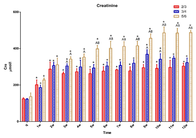
Fig. 7
CRE in 15 pigs (n = 5 for ⅔ group, n = 5 for ¾ group, n = 5 for ⅚ group) q – preoperative baseline
* p < 0.05 compared with the preoperative baseline. A p < 0.05 compared with value of ⅔ group at the same time point. B p < 0.05 compared with value of ¾ group at the same time point
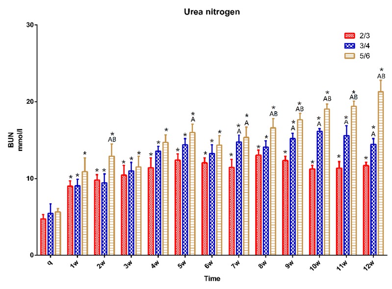
Fig. 8
BUN in 15 pigs (n = 5 for ⅔ group, n = 5 for ¾ group, n = 5 for ⅚ group) q – preoperative baseline
* p < 0.05 compared with the preoperative baseline. A p < 0.05 compared with value of ⅔ group at the same time point. B p < 0.05 compared with value of ¾ group at the same time point
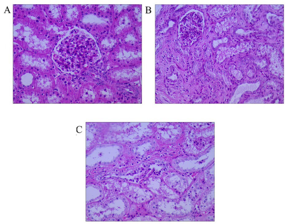
Fig. 9
Histological examination after ⅚ nephrectomy. The picture was captured in microscope vision.
A – normal renal tissue, B – at 4 weeks after surgery, C – at 10 weeks after surgery. All at 400×