
Table 1
List of primers used for amplification of dengue virus NS3 gene
| No. | Primer Name | Primer Sequences | Primer position within N2L1 sequence |
|---|---|---|---|
| 1 | NS3-1-OF | CAGGACTTTTCCCCGTATCA | 4419-4438 |
| 2 | NS3-1-IF | AACAACGGGCTGGAGTATTG | 4482-4501 |
| 3 | NS3-1-SEQ-F | CCAGGTCTTGGCATTAGAGC | 4774-4793 |
| 4 | NS3-2-OF | TCACAGACCCAGCAAGCATA | 5352-5371 |
| 5 | NS3-2-IF | AGCAGAGACCCATTTCCTCA | 5450-5469 |
| 6 | NS3-2-SEQ-IF | GGCAGAAATGGGTGCTAACT | 5719-5738 |
| 7 | NS3-1-OR | GGAACTCTGGACATGAGTGGGT | 5541-5520 |
| 8 | NS3-1-IR | TTGAGGAAATGGGTCTCTGC | 5451-5470 |
| 9 | NS3-2-OR | AGCATGATTGTACGCCCTTC | 6456-6475 |
| 10 | NS3-2-IR | TGGAAGCCTACCCATTTCTG | 6366-6385 |
Table 2
List of bacterial strains used in the present study
| No. | Bacterial strain | Genotype and description | Source |
|---|---|---|---|
| 1 | E. coli DH 5 α | F’, φ80d/lacZ.M15, recA1, endA1, gyrA96, thi-1, hsdR17(rK-, mK+), supE44, relA1, deoR, .(lacZYAargF) U169; | CEMB culture Collection |
| 2 | E. coli DH 5 α top10 | F- mcrA .(mrr-hsdRMS-mcrBC) ö80lacZ.M15 .lacX74 recA1 araD139 | CEMB culture Collection |
Table 3
Primers used for amplification and expression of dengue virus NS3 gene. Restriction sites were added at the start of primers
| No. | Genes | Primer sequence |
|---|---|---|
| 1 | NS3F-1 | GCAAGCTTGCCATGGCCGCTGGAGTATTGTCGGC |
| 2 | NS3F-2 | GCGGATCCGCCATGGCCGCTGTATTGTCGGC |
| 3 | NS3R | GCGCGGCCGCTTTCTTCCACTGCAAACTCTTTGTTC |
Table 4
pcDNA3.1 vector specific primer sequences
| Name | Primer sequence |
|---|---|
| T7 | TAATACGACTCACTATAGGG |
| BGH | TAGAAGGCACAGTCGAGG |
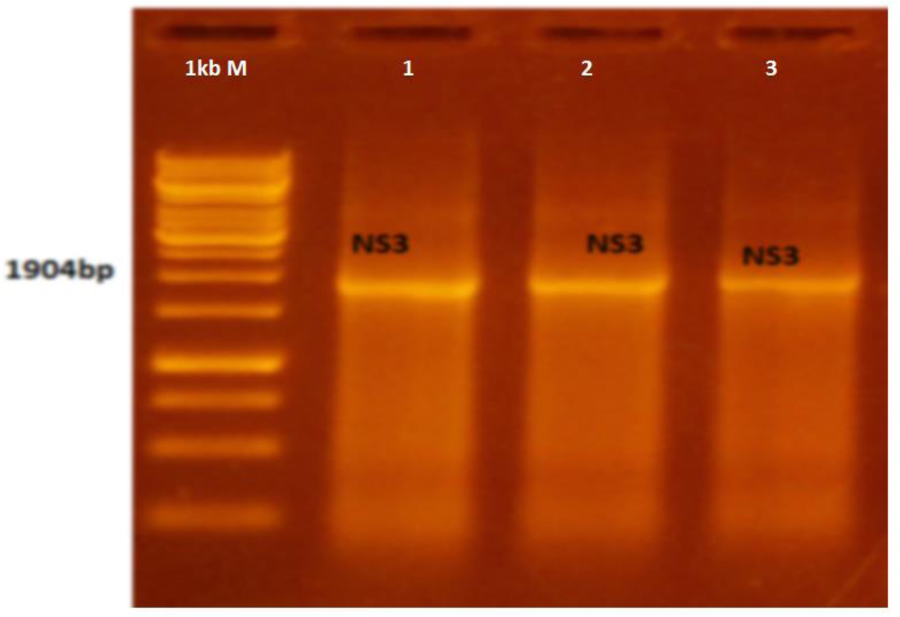
Fig. 1
PCR amplification of dengue NS3 gene. Lane M – 1Kb marker; lanes 1-3 – amplified NS3 product
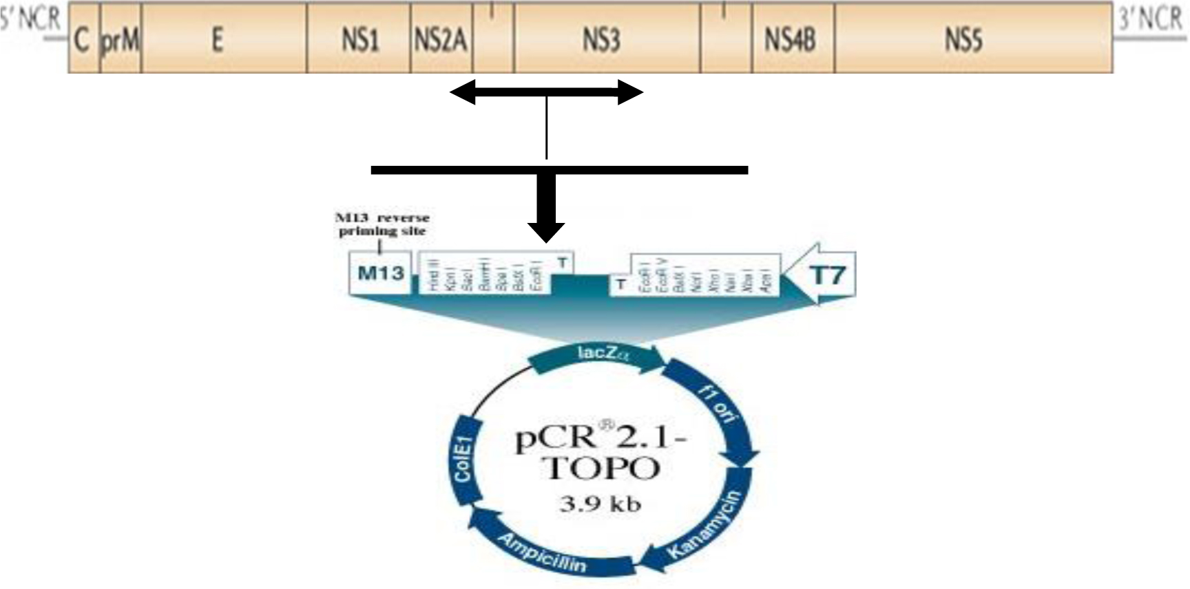
Fig. 2
The cloning of fragment containing NS3 region in pCR 2.1 TOPO
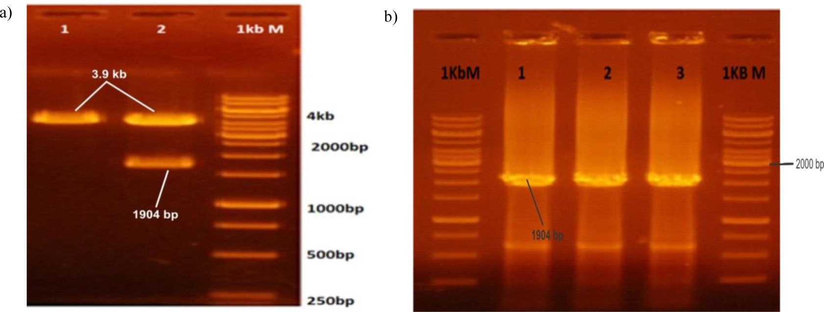
Fig. 3
(a) Restriction and digestion of NS3 encoding TA vectors. b) Cloning PCR
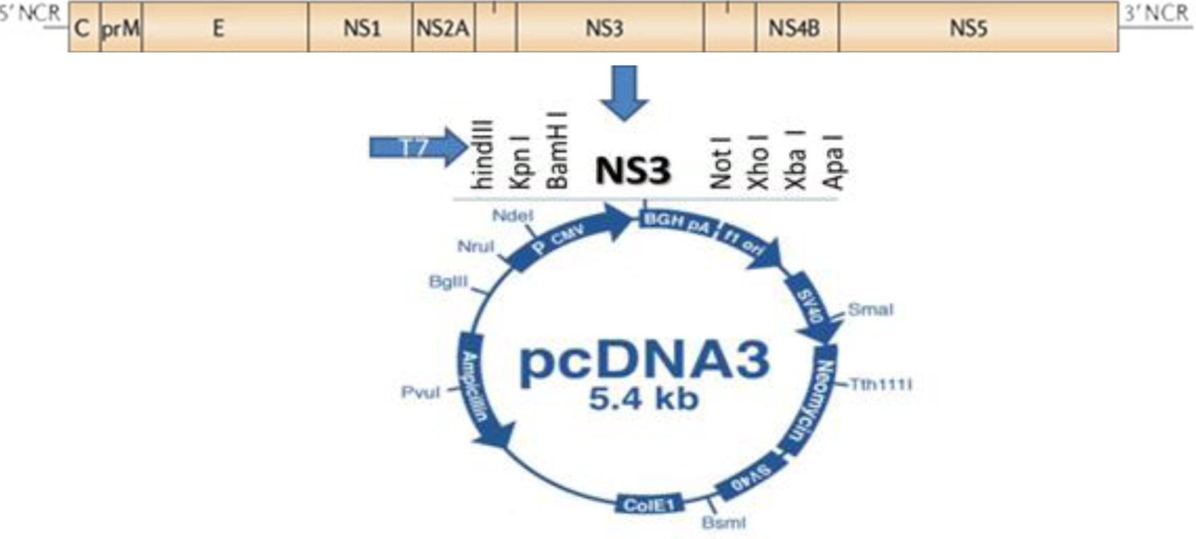
Fig. 4
Map of pcDNA3.1 mammalian expression vector
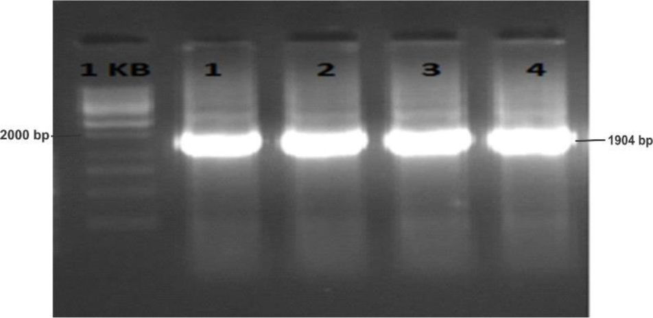
Fig. 5
PCR amplification of NS 3 gene with restriction site specific primers. Lane 1 M – 1 kb marker, lanes 2–4 – NS3 (1,904 bp)
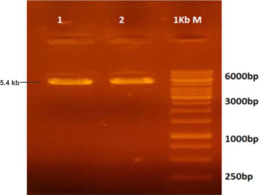
Fig. 6
Double-digested pcDNA3.1 mammalian expression vector. Lane M – 1kb marker; lanes 1 and 2 – digested pcDNA 3.1
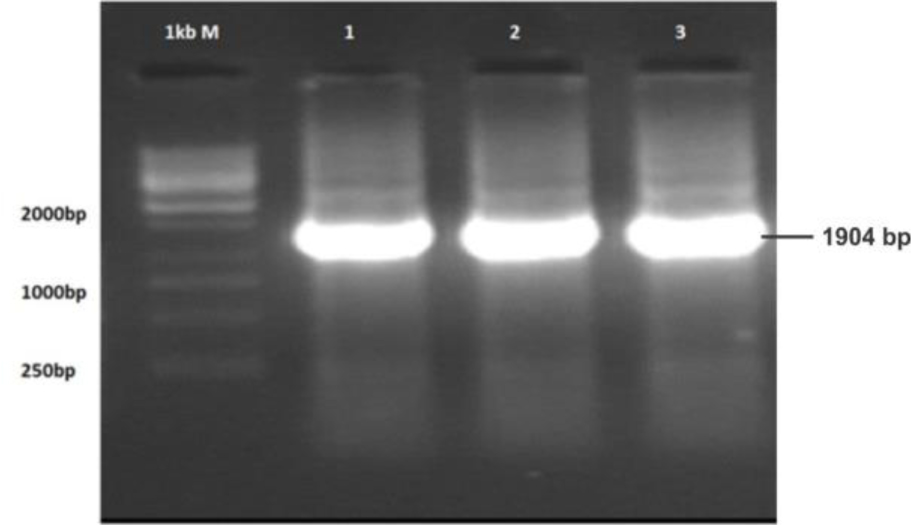
Fig. 7
Double-digested amplified NS3 gene of dengue virus. Lane M – 1kb marker; lanes 1–3 – double digested NS3 gene
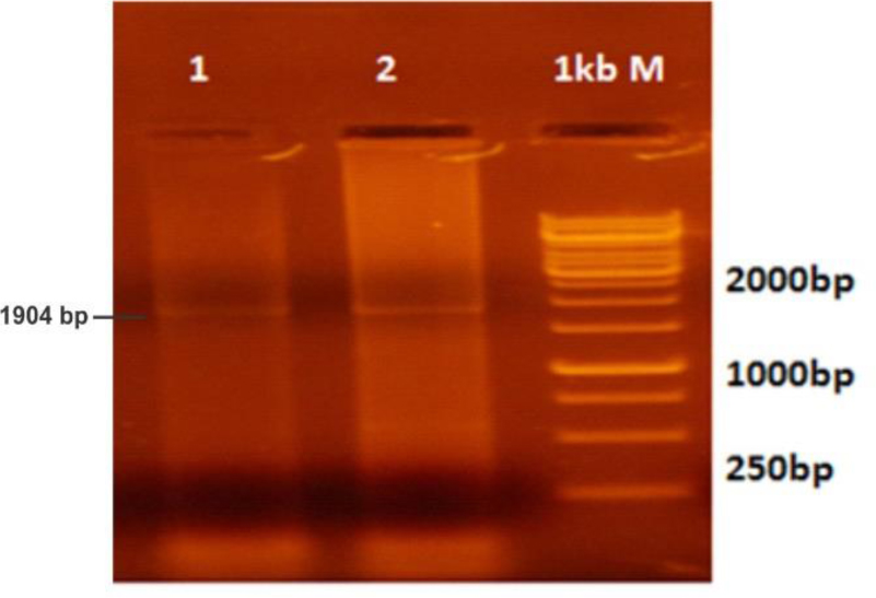
Fig. 8
Colony PCR (gene-specific) screening of NS3 encoding region in mammalian expression vector. Lanes 1–2 – positive NS3 encoding clones; lane M – 1kb ladder
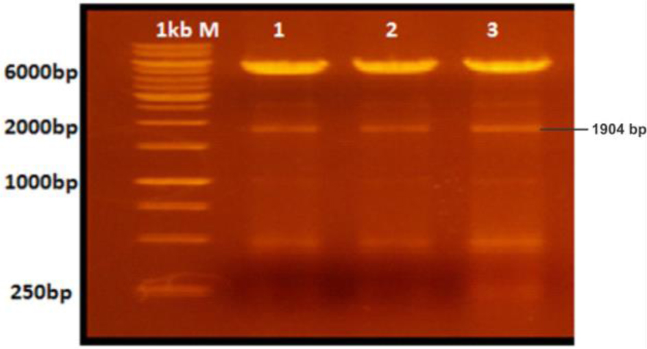
Fig. 9
Restriction and digestion of NS-3 encoding mammalian expression vector. Lane M – 1kb marker; lanes 1–3 – positively digested plasmids
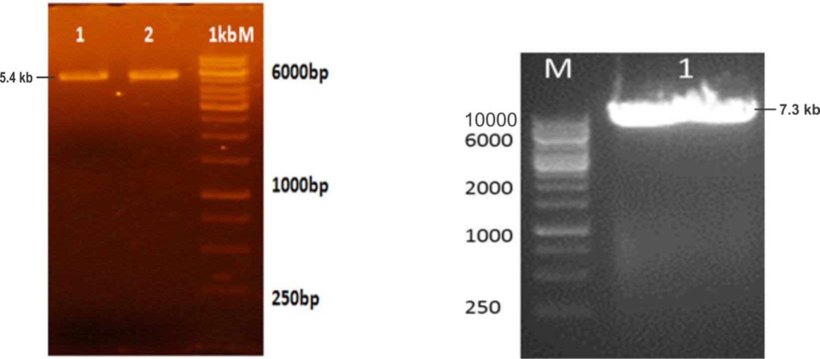
Fig. 10
pcDNA3.1 BglII linearised plasmid and empty vector. Lane M – 1kbM ladder; lanes 1 and 2 – linearised pcDNA3.1; lane M – 1kb ladder; lane 1 – linearised pcDNA3.1/NS3
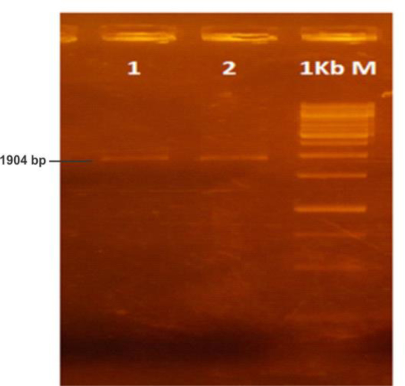
Fig. 11
NS3 expressing cell line characterised by RT-PCR