
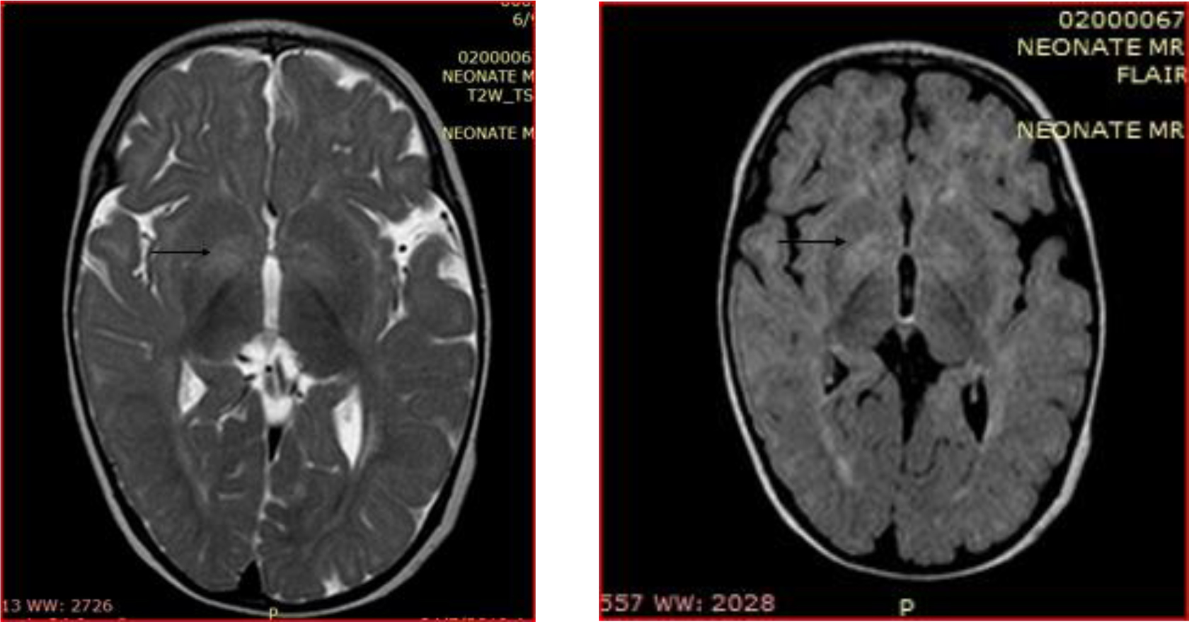
Figure 1.
Magnetic resonance imaging of the brain. T2-weighted and FLAIR sequences with symmetrical hyperintensities of globus pallidi mainly in the anterior part (arrowhead) and delayed progress of the myelination with regards to the patient’s age.
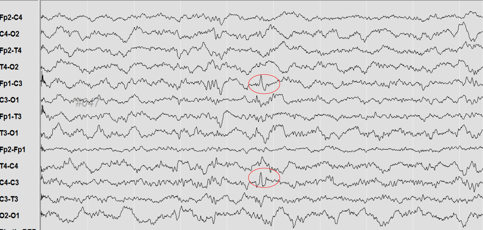
Figure 2.
EEG at the age of diagnosis, showed low voltage spikes mainly in the frontal area.
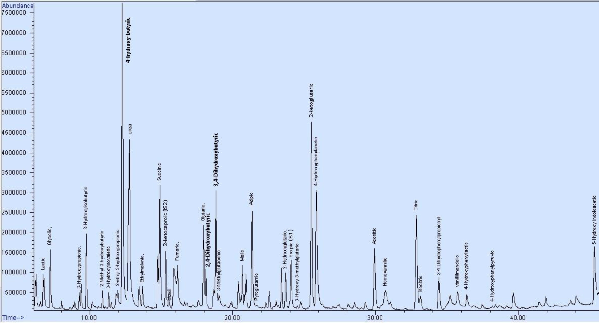
Figure 3.
Chromatogram of urine organic acids of the patient with SSADH, by Gas Chromatography-Mass Spectrometry. The presence of the diagnostic metabolites 4-hydroxy-butyic, 3,4-dihydroxybutyric acid and 2,4-dihydroxybutyric acid confirm the characteristic pattern of the disease.
Table 1.
Values of the metabolites of the child with SSADH. Pathological values are shown in bold
| Metabolite | Biological fluid/analysis | Value | Reference value |
| 4-hydroxy-butyric | Urine/Organic acids | 688.7 mmol/mol creatinine | n.d.1 |
| 3,4- dihydroxybutyric | Urine/Organic acids | 180 mmol/mol creatinine | n.d.1 |
| 2,4-dihydroxybutyric | Urine/Organic acids | 30 mmol/mol creatinine | n.d.1 |
| 3-hydroxy-propionic | Urine/Organic acids | 149.4 mmol/mol creatinine | 1.0–36.0 mmol/mol creatinine |
| 2-hydroxy-glutaric2 | Urine/Organic acids | 76.8 mmol/mol creatinine | 0.0–15.0 mmol/mol creatinine |
| glutaric2 | Urine/Organic acids | 36.2 mmol/mol creatinine | 0.0–5.3 mmol/mol creatinine |
| glycine | Plasma/Aminoacids CSF/Aminoacids | 289.0 μmol/L 4.0 μmol/L | 107.0–343 μmol/L 3.7–7.6 μmol/L |
| 5-hydroxy-indoloacetic | CSF/ Biogenic amines | 184.0 nmol/L | 170.0–490.0 nmol/L |
| homovanillic | CSF/ Biogenic amines | 630.0 nmol/L | 344.0–906.0 nmol/L |
| 3-ortho-methyldopa | CSF/ Biogenic amines | 20.0 nmol/L | 4.0–50.0 nmol/L |
| 5-hydroxy-tryptophan | CSF/ Biogenic amines | 7.0 nmol/L | 1.6–15.0 nmol/L |
| neopterin | CSF/ Pterins | 31.0 | 8.0–43.0 nmol/L |
| biopterin | CSF/ Pterins | 41.0 | 8.0–54.0 nmol/L |
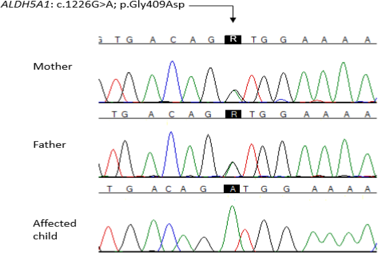
Figure 4.
Electropherograms depicting the parental heterozygosity as well as the patient’s homozygosity for the pathogenic variant.
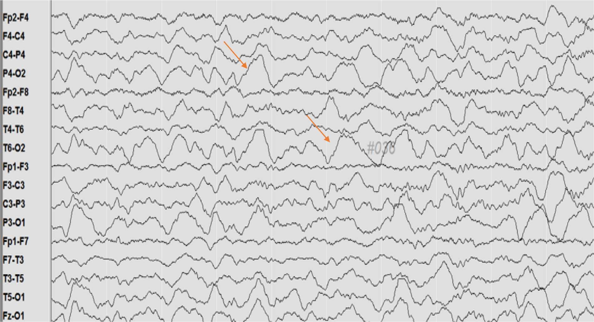
Figure 5.
Interictal sleep EEG at the age of 2 ½ years old, with intermittent rhythmic delta activity (1–2Hz, up to 250 μV) mainly in parietal-occipital area bilaterally.
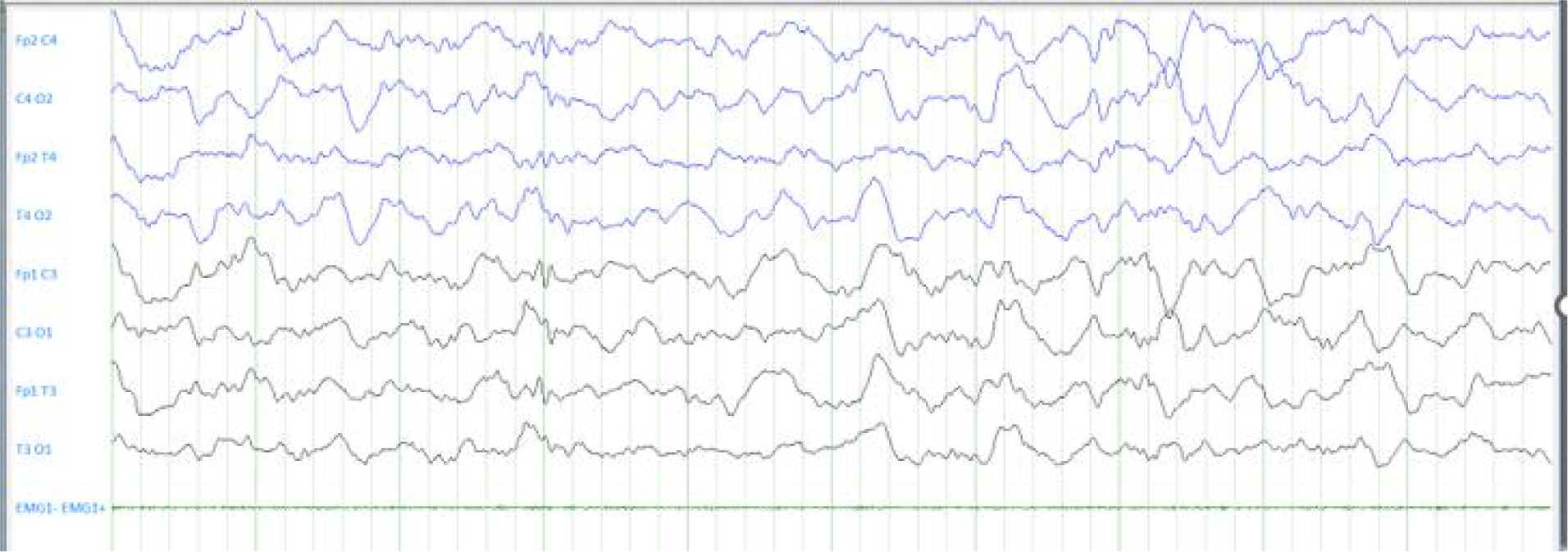
Figure 6.
Interictal sleep EEG, at the age of 4 years old, shows low voltage spikes mainly in frontal-temporal area bilaterally.