
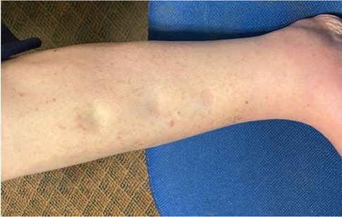
Figure 1
Extent of pitting edema in calf prior to DTM.
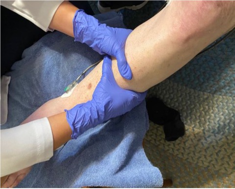
Figure 2
Position of hands during DTM.
| 0 | start treatment session - placed leg on chair |
| 15 | start massage and end of instrumentation period |
| 20 | stop massage - start recovery |
| 60 | placed leg on floor |
| 120 | end of treatment session |
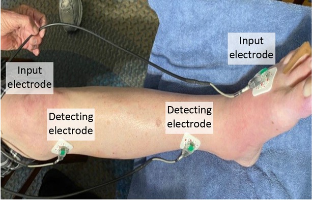
Figure 3
EIS electrode placement on the calf.
Table 1
Vblood, Vcell, Vinstl Volumes and Changes in Volume (all mL) at Elapsed Times (min.) of changes in the DTM treatment.
| ELAPSE TIME - Min. | Vblood | Change in Vblood | Vcell | Change in Vcell | Vinstl | Change in Vinstl |
|---|---|---|---|---|---|---|
| 0 | 1035 | 0 | 926 | 0 | 1067 | 0 |
| 15 | 998 | -37 | 907 | -19 | 1054 | -13 |
| 20 | 1013 | 15 | 816 | -91 | 994 | -60 |
| 60 | 1006 | -7 | 556 | -260 | 1001 | 7 |
| 120 | 957 | -49 | 702 | 146 | 1033 | 31 |
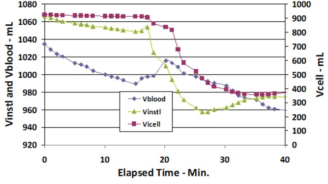
Figure 4
Vblood, Vinst) and Vcell volumes (mL) of the calf during start of DTM vs. Elapsed Time (min).
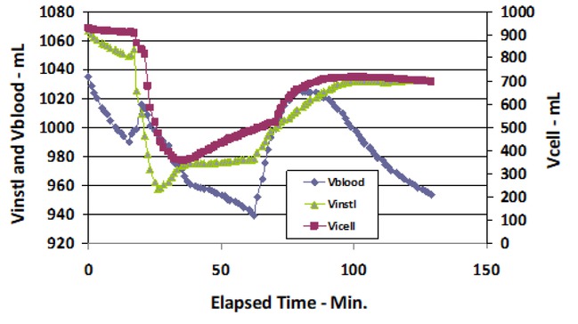
Figure 5
Vblood, Vinst and Vcell compartment volumes (mL) of the calf during the total DTM treatment and recovery periods vs. Elapsed Time (min).