
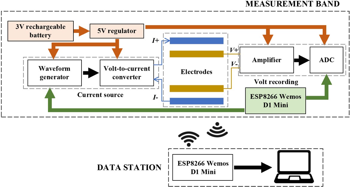
Fig. 1
The overall architecture of the system.
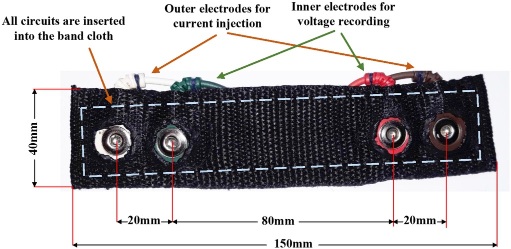
Fig. 2
The band prototype of 150mm x 40mm in dimension and the distances between.
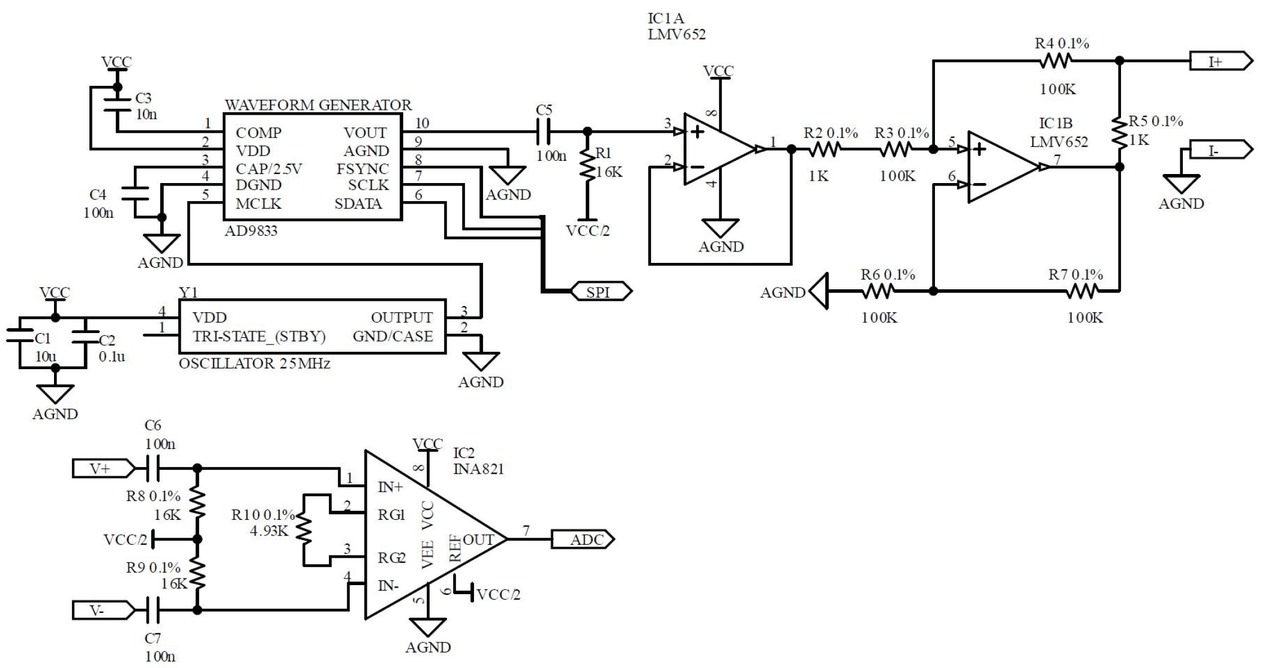
Fig. 3
The schematic of the current source and voltage recording on the band
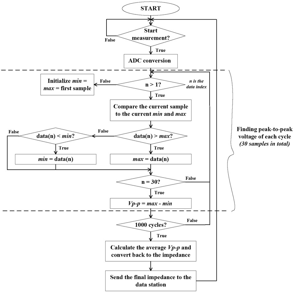
Fig. 4
Flowchart of the signal processing
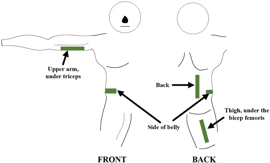
Fig. 5
Four locations were picked for the local impedance measurement: on the upper arm under the triceps, on the side of the belly, on the side of the back and on the thigh under the bicep femoris
Table 1
The information of volunteers in the experiment including age, BME, body fat (%) and fasting glucose
| Age | BMI | Body fat (%) | Fasting glucose (mmol/l) | |
|---|---|---|---|---|
| mean ± | 31.9 ± | 22.1 ± | 20.7 ± 6.9 | 7.4 ± |
| std | 6.6 | 3.5 | 1.8 | |
| min | 18 | 17.3 | 6.2 | 4.9 |
| max | 44 | 27.6 | 33.1 | 10.9 |
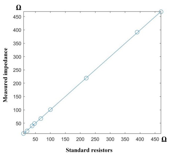
Fig. 6
Results between the impedance measured by the band and the standard resistors
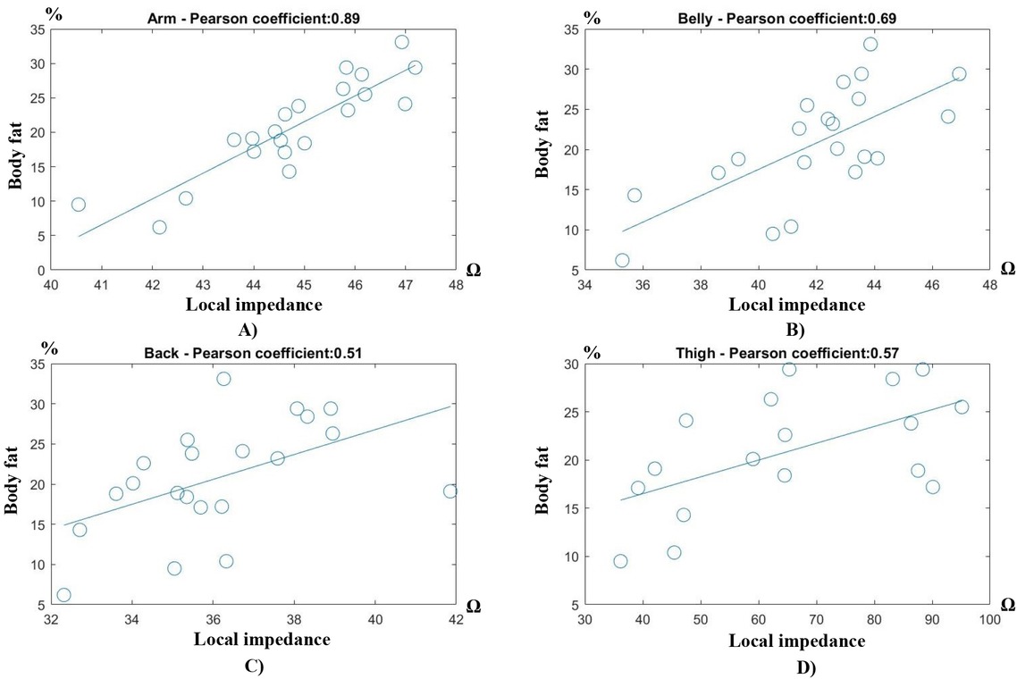
Fig. 7
Local impedance versus body fat at 4 locations: A) upper arm, B) belly, C) back, and D) thigh.
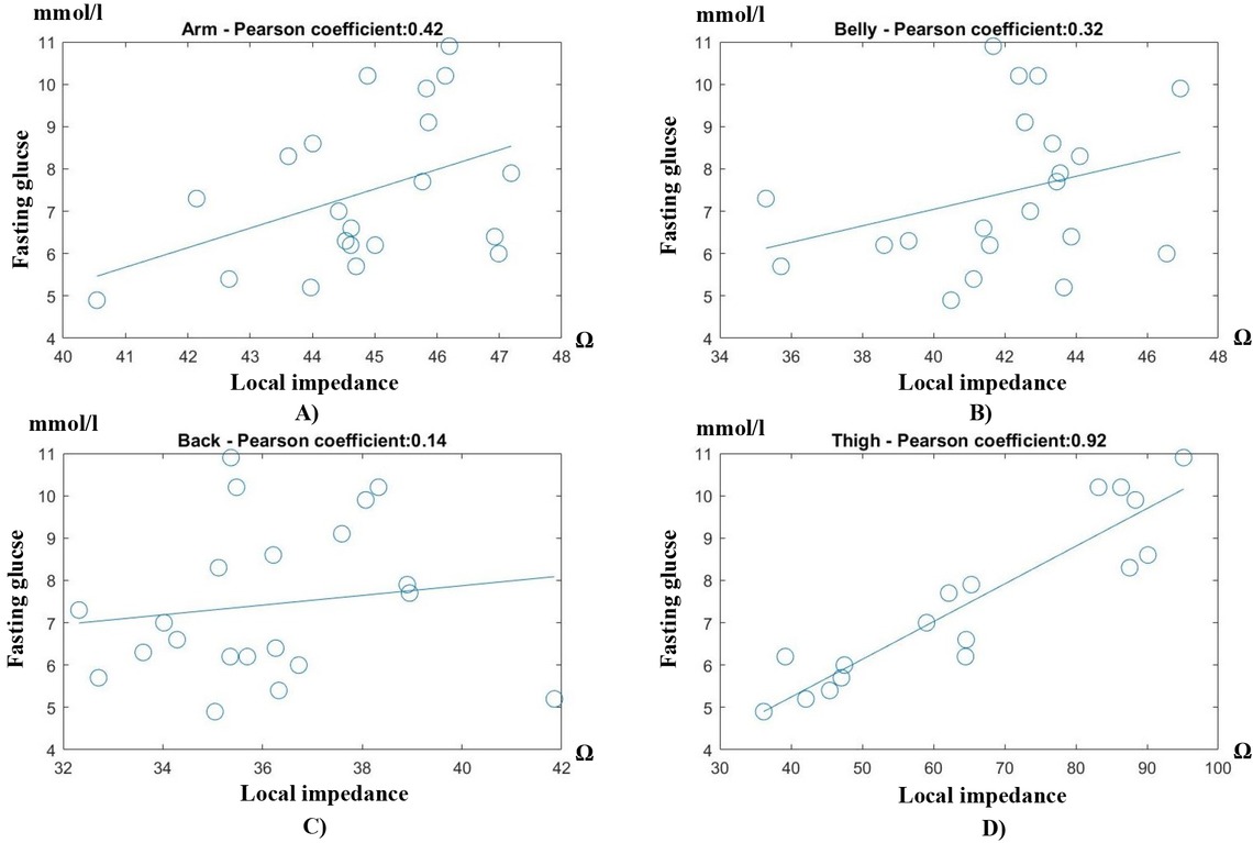
Fig. 8
Local impedance versus fasting glucose level at 4 locations: A) upper arm, B) belly, C) back, and D) thigh.
Table 2
Correlation coefficients and the corresponding confidence intervals (95%) between the local impedances measured at 4 locations: the upper arm, the belly, the back and the thigh, and the two parameters: the body fat and fasting glucose of all 21 volunteers. The best results are highlighted in boldness and red. Abbreviation: Cor. Coef: correlation coefficient. CI: confidence interval (95%)
| Arm | Belly | Back | Thigh | |||||
|---|---|---|---|---|---|---|---|---|
| Cor. Coef.* | CI** | Cor. Coef.* | CI** | Cor. Coef.* | CI** | Cor. Coef.* | CI** | |
| Body fat | 0.89 | 0.73-0.95 | 0.69 | 0.36-0.86 | 0.51 | 0.09-0.77 | 0.57 | 0.13-0.82 |
| Fasting Glucose | 0.42 | -0.02-0.72 | 0.32 | -0.14-0.65 | 0.14 | -0.31-0.54 | 0.92 | 0.81-0.97 |