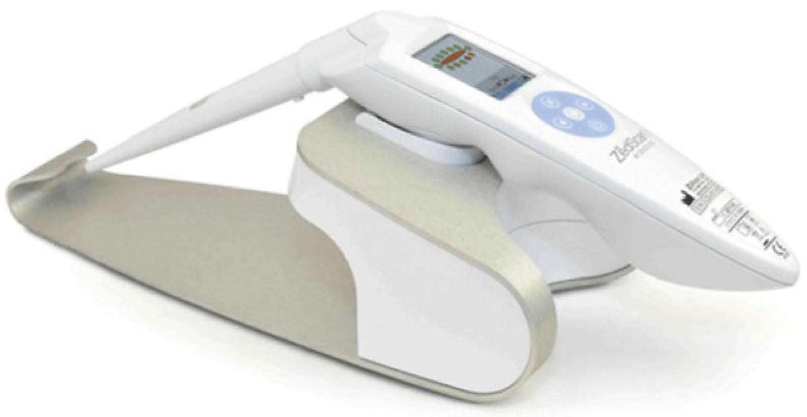
Have a personal or library account? Click to login

Prognostic value of Electrical Impedance Spectroscopy (EIS) when used as an adjunct to Colposcopy – a longitudinal study

Open Access
|Nov 2020

Figures & Tables

Figure 1

The ZedScanTM handset, used to make the EIS measurements. The handset is shown placed on the base. The tapered disposable is shown on the left and terminates in the four AgCl electrodes that are placed on the tissue. The screen is used to guide the clinician to take up to 12 measurements around the transformation zone of the cervix.

Figure 2

This shows the median of all the 31 spectra where CIN2+ occurred in black with the 95% confidence levels shown in red. The median of all the 536 spectra where CIN2+ did not occur is also shown in black, but with the 95% confidence levels shown in blue. Log scales are used on both axes.

Figure 3

An ROC curve comparing the EIS impedance of the two groups, at a frequency of 152 Hz, who either did or did not develop CIN2+. The AUC is 0.621, p=0.0088 when using a Mann-Whitney U test.

Figure 4

Comparison of the frequency differential of the impedance spectra for the CIN2+ women (upper curve) and the <CIN2+ women (lower curve). The curves are from the fitted Cole equation. The parameters of the equation are given in Table 3.

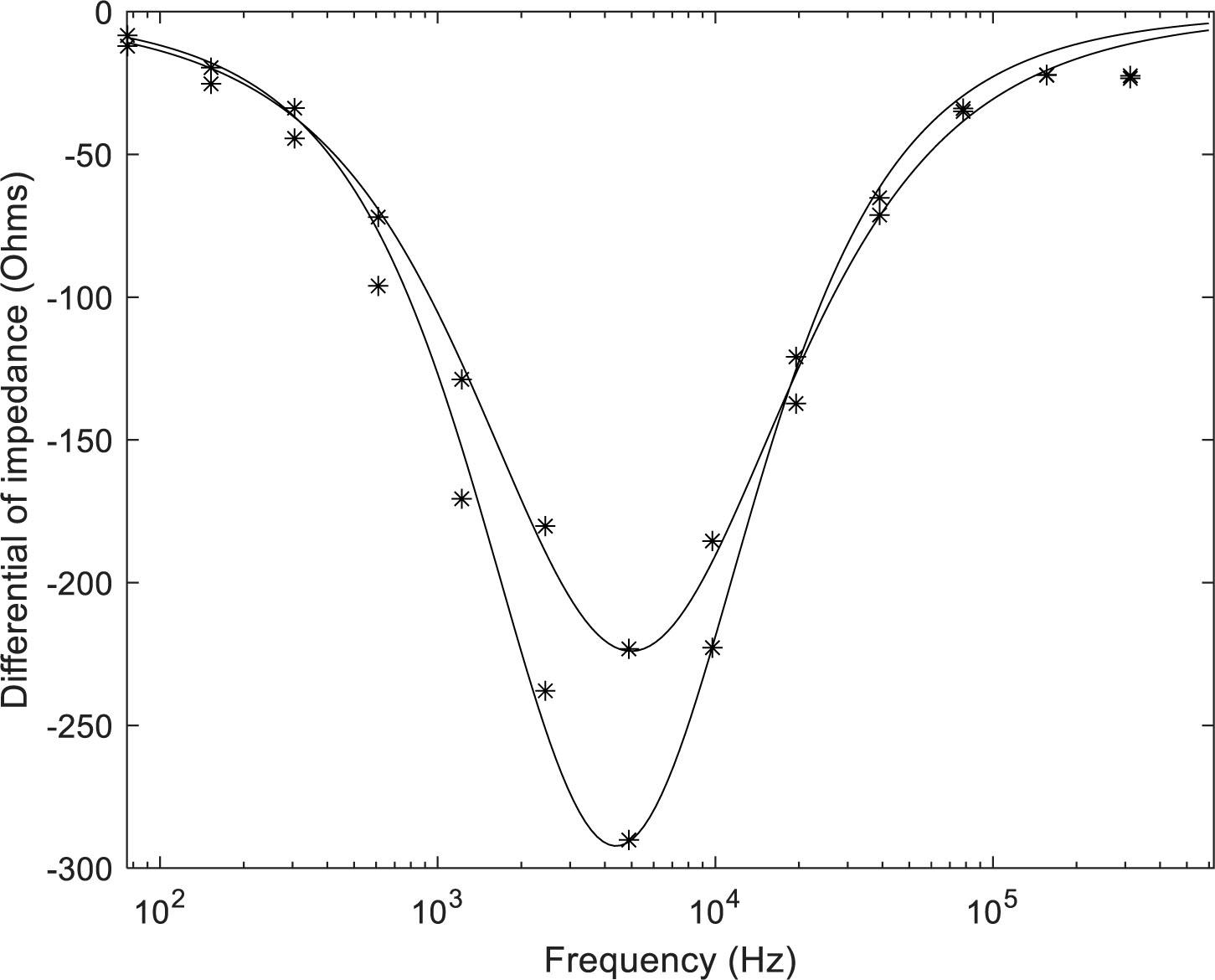
The Cole equation parameters of the curves shown in Fig 1_

Women who did not develop CIN2+Ro = 1485R = 80Fc = 4500α = 0.18
Women who did develop CIN2+Ro = 1220R = 90Fc = 5100α = 0.24

This shows how the 847 women were grouped for analysis_ The prevalence of CIN2+, and the annual risk of disease, is also given for each group_ The annual risk takes into account the fact that the first three groups of women did not have the same median period from the time of the index colposcopy to the time when disease was detected_

Outcome of colposcopy (CI) and EISPrevalence of CIN2+ within 3 yearsPercentage risk of developing CIN2+ per annum, over the follow-up period for each woman
Group 1396 womenBoth CI and EIS negative(Double negative)CIN2+ arose in 13 (3.28%)1.20%
Group 2318 womenCI negative but EIS positiveCIN2+ arose in 12 (3.77%)1.64%
Group 3123 womenBoth CI and EIS positive(Double positive)CIN2+ arose in 10 (8.13%)4.57%
Group 410 womenCI positive but EIS negativeNo cases of CIN2+N/A

The breakdown of the prevalence of disease in women where the index CI was negative and at least one EIS point was above the higher probability index threshold of 0_87_

Number of abnormal EIS pointsNumber of womenCases of CIN2+ within three yearsPrevalence of disease
One22673.1%
Two11154.50%
Three or more104109.62%
One or two337123.56%
Language: English
Page range: 81 - 86
Submitted on: Aug 25, 2020
Published on: Nov 6, 2020
Published by: University of Oslo
In partnership with: Paradigm Publishing Services
Publication frequency: 1 times per year

© 2020 B. H. Brown, P.E. Highfield, J. A. Tidy, published by University of Oslo
This work is licensed under the Creative Commons Attribution 4.0 License.