
Table 1
Overview of extracted parameters from the EDA responses that were used in the data analyses.
| Parameter | Description | Unit |
|---|---|---|
| SCR_Amp | Amplitude of the skin conductance response | μS |
| SPR_Amp | Amplitude of the skin potential response | mV |
| SSR_Amp | Amplitude of the skin susceptance response | μS |
| SPRET | Turning point of the SPR relative to the SCR peak | % |
| SCR_Trise | Time from onset of SCR to peak SCR | sec. |
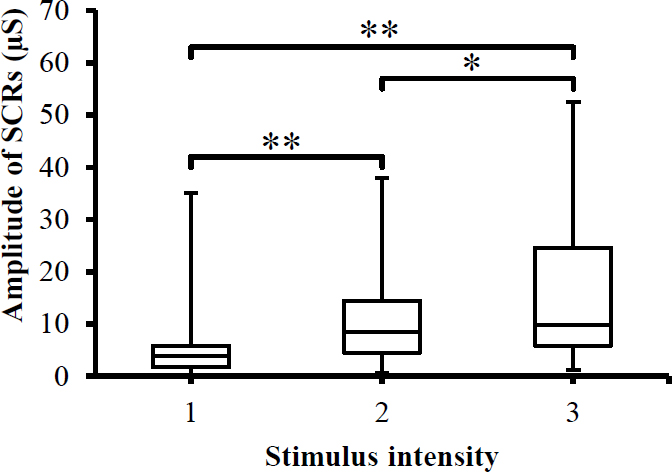
Fig. 1
Box-plot with medians, quartiles and the min and max as whiskers shows value of SCRs_Amp with respect to the stimulus intensity. *p<0.05 and **p<0.005.
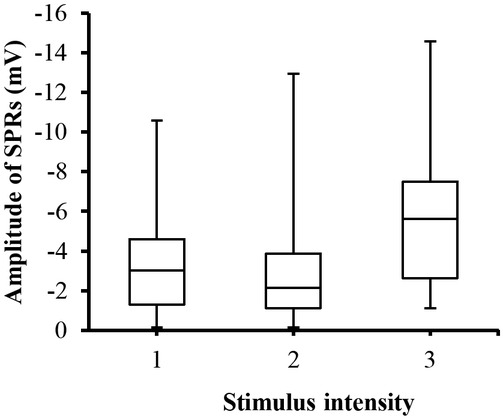
Fig. 2
Box-plot with medians, quartiles and the min and max as whiskers shows value of SPRs_Amp with respect to the stimulus intensity.
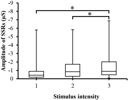
Fig. 3
Box-plot with medians, quartiles and the min and max as whiskers shows value of SSRs_Amp with respect to the stimulus intensity. *p<0.05.
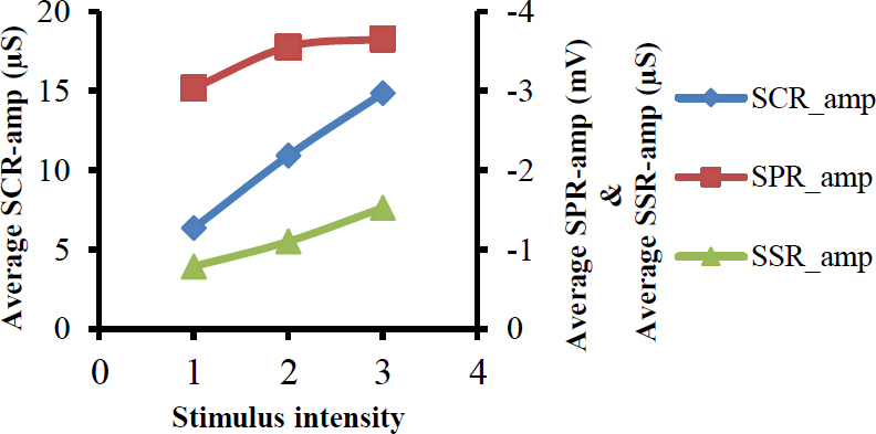
Fig. 4
Shows relation between average values of amplitudes of SCRs, SPRs and SSRs on one side, and the stimulus intensity on other side.
Table 2
Statistical analysis with a linear mixed effects model.
| 95% CI | ||||
|---|---|---|---|---|
| Parameters | Estimate | p value | Lower | Upper |
| SCRs | 4.255 | <0.001 | 2.490 | 6.019 |
| SPRs | -0.296 | 0.157 | -0.709 | 0.116 |
| SSRs | -0.384 | 0.001 | -0.620 | -0.143 |
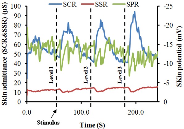
Fig. 5
A typical EDA recording from a participant, demonstrating changes in EDA responses with respect to painful stimuli.
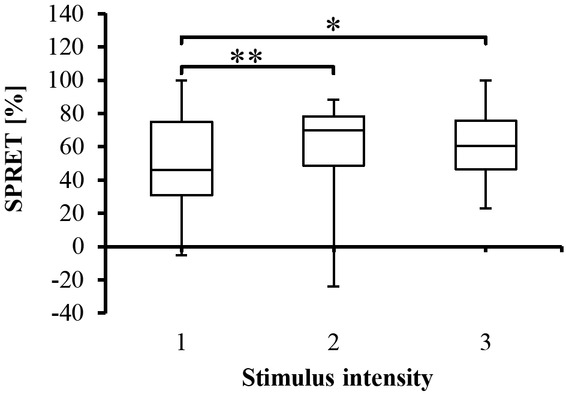
Fig. 6
Box-plot with medians, quartiles and the min and max as whiskers, showing the SPRET percentage for all participants with respect to the stimulus intensity.*p<0.05 and **p<0.005.
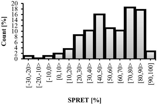
Fig. 7
SPRET percentage for each category.
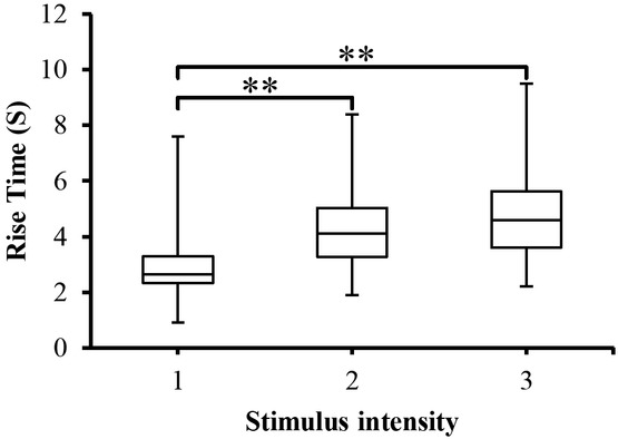
Fig. 8
SCR_Trise to the different stimulus intensity presented as box-plots with medians, quartiles and the min and max as whiskers. **p<0.005.