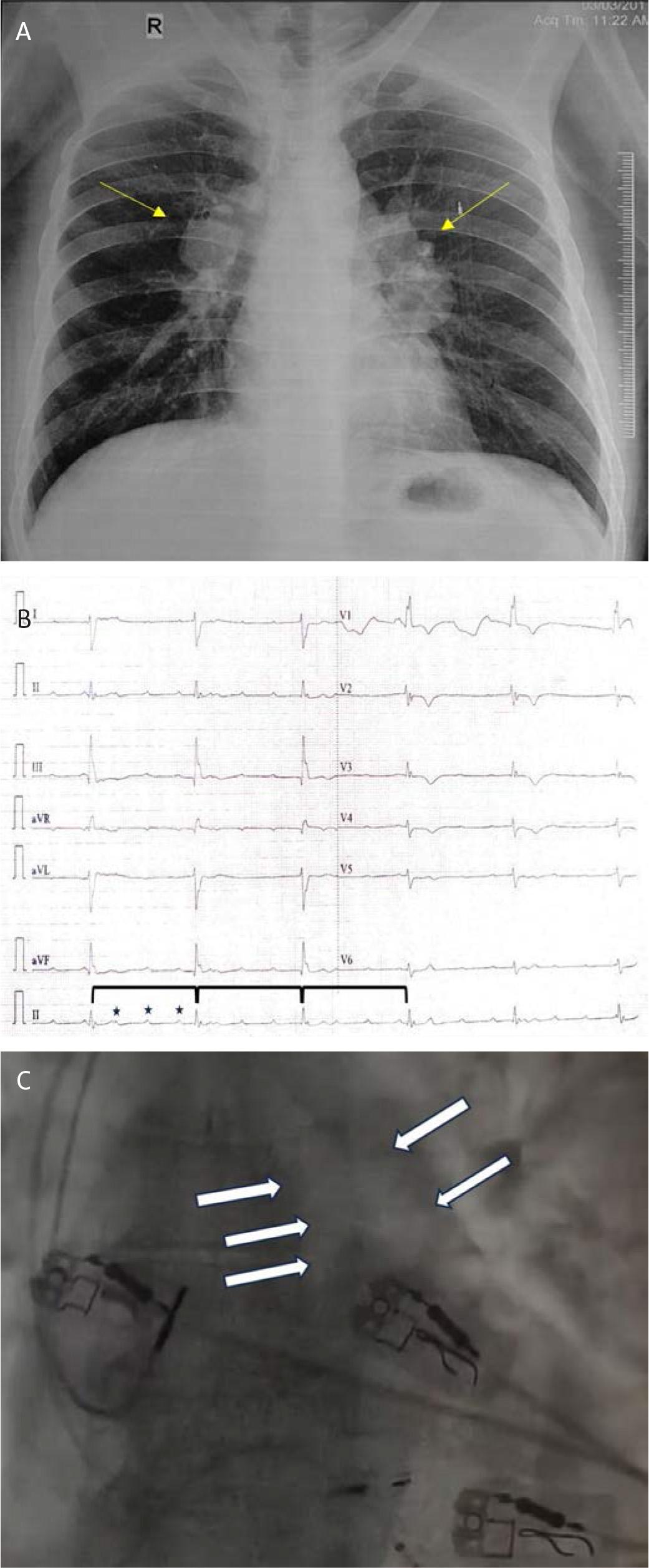
Have a personal or library account? Click to login
Air Lock Syndrome – an Unusual Complication of Pacemaker Implantation Cover

Air Lock Syndrome – an Unusual Complication of Pacemaker Implantation

Open Access
|Mar 2024

Figures & Tables

FIGURE 1.

A, Conventional X-ray, PA view, showing bilateral hilar adenopathy (yellow arrow). B, 12-lead electrocardiogram showing complete dissociation of p waves (black stars) and QRS escape rhythm (black bar). C, Fluoroscopic image showing clear delineation of the RVOT and pulmonary tract (white arrow), which is normally not visible in fluoroscopy.
DOI: https://doi.org/10.2478/jce-2024-0005 | Journal eISSN: 2457-5518 | Journal ISSN: 2457-550X
Language: English
Page range: 45 - 47
Submitted on: Jan 3, 2024
Accepted on: Feb 6, 2024
Published on: Mar 21, 2024
Published by: Asociatia Transilvana de Terapie Transvasculara si Transplant KARDIOMED
In partnership with: Paradigm Publishing Services
Publication frequency: 4 issues per year

© 2024 Soorampally Vijay, Bharath Raj Kidambi, Sriram Veeraraghavan, published by Asociatia Transilvana de Terapie Transvasculara si Transplant KARDIOMED
This work is licensed under the Creative Commons Attribution-NonCommercial-NoDerivatives 3.0 License.