Fig. 1.
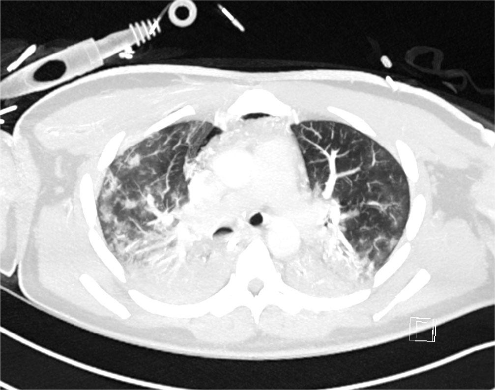
Fig. 2.
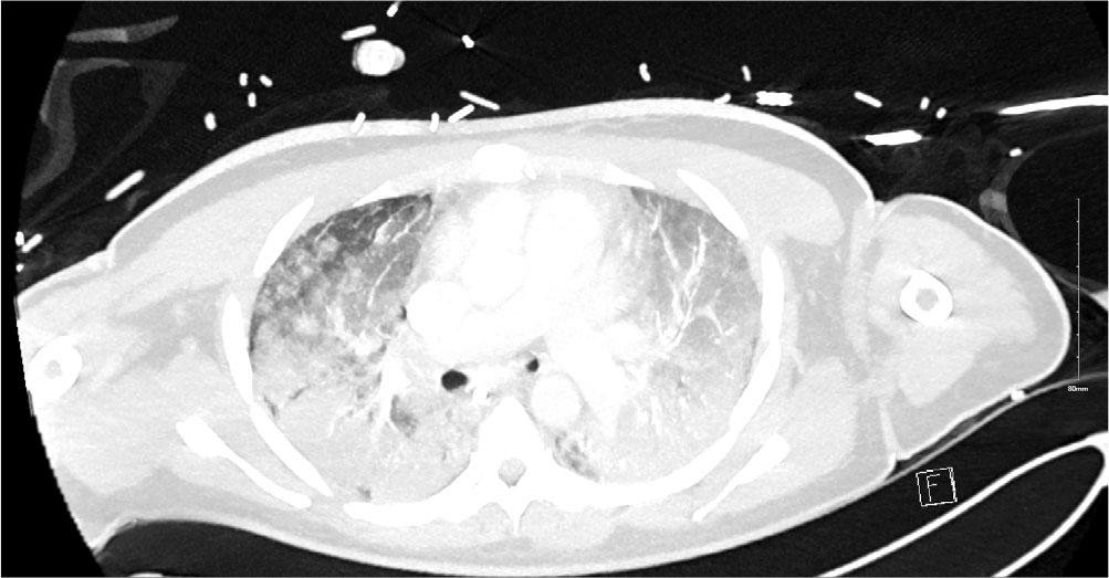
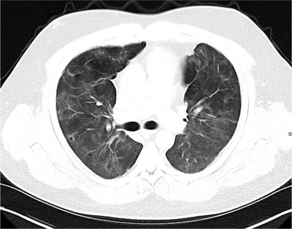
Fig. 3.
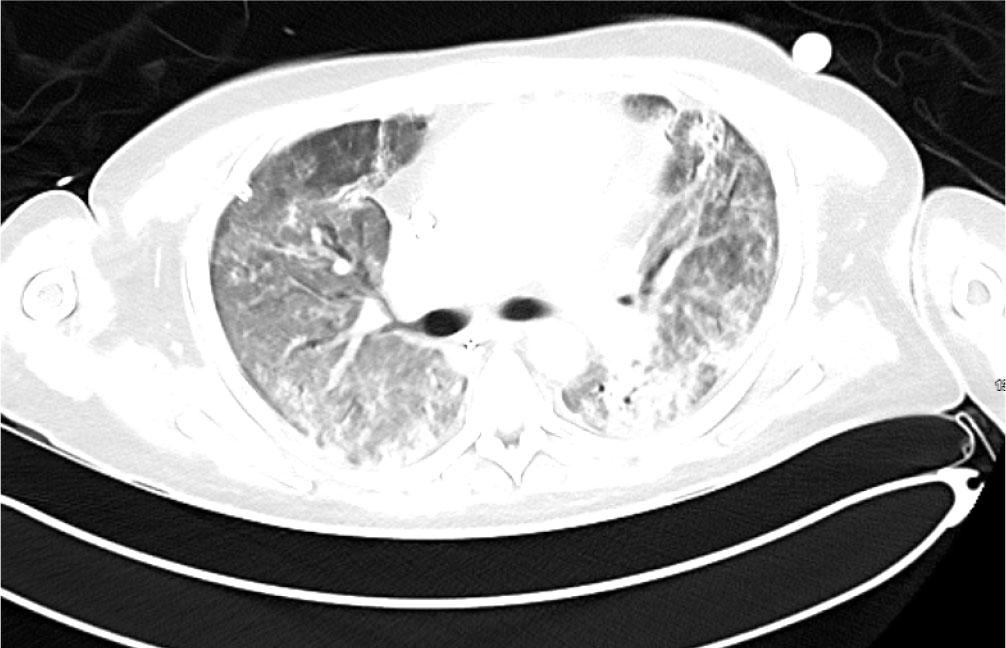
Fig. 4.
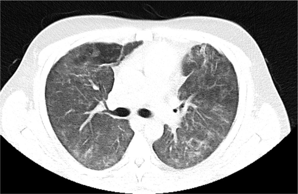
Fig. 5.

© 2026 Samreen Tariq, Fiona O’Hannigan, Nizrull Nasir, Serena O’Brien, Brian Marsh, Jennifer Hastings, John Stack, Josephine Kelliher, Katherine O’Reilly, published by University of Medicine, Pharmacy, Science and Technology of Targu Mures
This work is licensed under the Creative Commons Attribution 4.0 License.