
Table 1.
Arterial Blood Gas (ABG) Trends Demonstrating Severe Metabolic Acidosis Following HoLEP and Resolution Post-CRRT.
| ABG parameter | Intraop | Day 0 | Day 1 AM | Day 1 PM | Day 2 | Day 3 |
|---|---|---|---|---|---|---|
| FIO2 (%) | 1 | 0.5 | 0.3 | 0.3 | 0.3 | 0.21 |
| pH | 7.102 | 7.141 | 7.211 | 7.398 | 7.412 | 7.441 |
| pCO2 (mmHg) | 42.2 | 37.6 | 35.9 | 35.8 | 35.6 | 33.9 |
| pO2 (mmHg) | 278 | 151.6 | 79.4 | 82.8 | 98.9 | 78.6 |
| Base Excess (mEq/L) | −16 | −15.6 | −12.8 | −2.8 | −2.1 | −1.2 |
| Bicarbonate (mEq/L) | 13.2 | 12.7 | 14.5 | 20.6 | 22.6 | 23.4 |
| Sodium (Na+, mEq/L) | 141 | 136 | 139 | 138 | 139 | 139 |
| Potassium (K+, mEq/L) | 4.3 | 6.0 | 4.5 | 4.0 | 3.9 | 3.5 |
| Chloride (Cl−, mEq/L) | 115 | 115 | 113 | 109 | 110 | 105 |
| Lactate (mmol/L) | 3.3 | 3.2 | 4.8 | 2.7 | 1.7 | |
| Glucose (mmol/L) | 12.1 | 14.9 | 15.9 | 6.6 | 8 | 7.7 |
| Anion Gap (mEq/L) | 12.8 | 8.3 | 11.5 | 8.4 | 6.4 | 10.6 |

Fig. 1.
Timeline of Clinical Events in HoLEP Case. CTAP – Computed Tomography of Abdomen and Pelvis. SPC – Suprapubic Catheter.
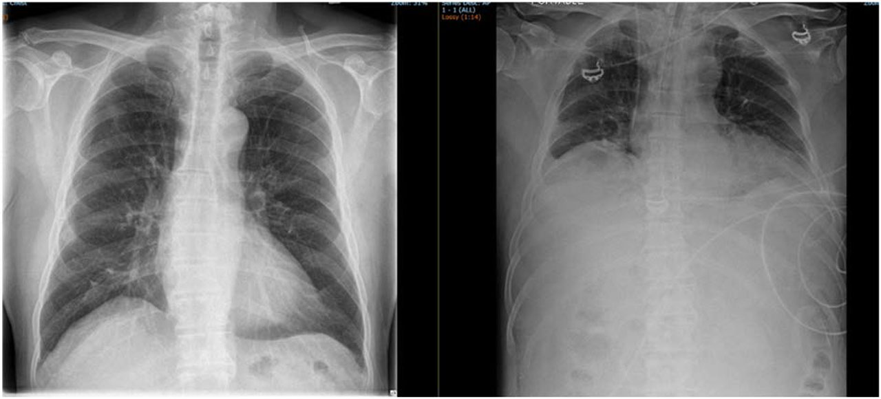
Fig. 2.
Comparison of Preoperative and Postoperative Chest X-rays in the patient with Abdominal Compartment Syndrome following HoLEP. The left image shows a normal preoperative chest X-ray, while the right image demonstrates postoperative abdominal distension and diaphragmatic splinting with reduced lung fields.
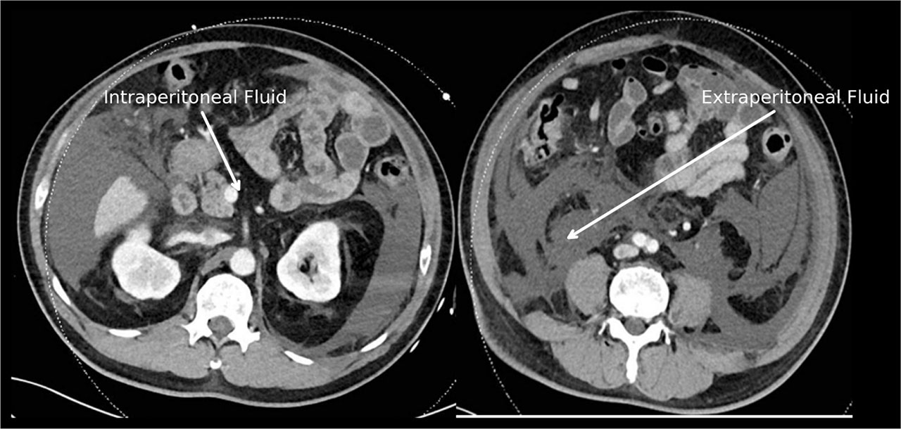
Fig. 3.
Abdominal CT done postoperatively on ICU admission showing fluid seepage into Extraperitoneal and Intraperitoneal Compartments. The left panel demonstrates intraperitoneal fluid (white arrow on the left) around the mesentery and bowel loops, while the right shows extraperitoneal fluid (white arrows on the right) in the retroperitoneal and perivesical spaces.