
Table 1.
Demographic data and complete blood count parameters in patients with and without sepsis-associated encephalopathy (SAE).
| Parameter | Patients with SAE (n=184) | Patients without SAE (n=96) | Reference ranges | P value |
|---|---|---|---|---|
| Age (years) | 73 (18) | 70 (16) | 0.088 | |
| Male sex | 94 (51.08%) | 52 (54.16%) | 0.624 | |
| Leucocyte count (per μL) | 14100 (10960) | 10265 (10882) | 4000–9000 | 0.003* |
| Hemoglobin (g/dL) | 10.1 (3.9) | 9.8 (3.8) | 11.5–17 | 0.276 |
| Platelet count (per μL) | 200000 (166500) | 128500 (149500) | 150000–400000 | <0.001* |
Table 2.
Multivariate logistic regression model using age and complete blood count parameters.
| Parameter | Adjusted OR | 95% CI | P value |
|---|---|---|---|
| Leucocyte count | 1.000 | 1.000; 1.000 | 0.403 |
| Platelet count | 1.003 | 1.001; 1.006 | 0.004* |
| Age | 1.003 | 0.987; 1.020 | 0.710 |
Table 3.
Hemostatic tests in patients with and without sepsis-associated encephalopathy (SAE).
| Parameter | Patients with SAE (n=63) | Patients without SAE (n=55) | Reference ranges | P value |
|---|---|---|---|---|
| PT (s) | 15.95 (3.8) | 17 (5.2) | 10.4–14.3 | 0.440 |
| Fibrinogen (mg/dL) | 431.88 (±198.14) | 373.94 (±170.94) | 200–400 | 0.095 |
| Factor II (%) | 57.46 (±15.12) | 57.07 (±14.73) | 70–120 | 0.888 |
| Factor V (%) | 54.35(±19.13) | 52.70 (±18.10) | 70–120 | 0.635 |
| Factor VII (%) | 36.33(±13.78) | 37.61(±15.54) | 55–165 | 0.638 |
| Factor X (%) | 56.34(±16.40) | 54.67(±15.62) | 70–120 | 0.579 |
| AT (%) | 65.44(±17.85) | 68.25(±14.45) | 80–120 | 0.389 |
| PC (%) | 58.98(±11.94) | 60.06(±13.71) | 70–150 | 0.663 |
| PS (%) | 44.86(±10.08) | 43.68(±12.04) | 60–140 | 0.581 |
| D-dimer (ng/mL) | 4830.5 (6273) | 3940 (7493) | <500 | 0.446 |
| EXTEM CT (s) | 69 (20.5) | 70.5 (22.8) | 38–79 | 0.791 |
| EXTEM CFT (s) | 90.5 (80) | 114.5 (104) | 34–159 | 0.001* |
| EXTEM MCF (mm) | 64.30(±14.41) | 56.71(±12.60) | 50–72 | 0.004* |
| EXTEM MCE | 218.81(±126.33) | 147.85(±73.31) | - | 0.001* |
| EXTEM CLI60 (%) | 98(3) | 98(4) | >85 | 0.167 |
| MaxVel (mm/min) | 17 (9.8) | 14 (10) | 11–25a | 0.021* |
| t-MaxVel (s) | 85 (69.5) | 99 (105.5) | 147–223a | 0.931 |
| AUC (mm x 100) | 6360.16(±1364.86) | 5602.31(±1269.07) | - | 0.005* |
| FIBTEM MCF (mm) | 21.71(±11.40) | 16.40(±7.78) | 9–25 | 0.007* |
| FIBTEM MCE | 38.44 (±53.24) | 20.66 (±13.23) | - | 0.024* |
| APTEM CLI60 (%) | 98 (3) | 98 (4) | - | 0.278 |
| Diff CLI60 | 0 (0) | 0 (1) | - | 0.374 |
| Platelet component | 175.5 (156.75) | 119.5 (105) | - | 0.010* |
INR, international normalized ratio; PT, prothrombin time; AT, antithrombin; PC, protein C; PS, protein S; AUC, area under the thrombus formation curve; CFT, clot formation time; CT, clotting time; EXTEM CLI60, lysis index at 60 min; MaxVel, maximum velocity of clot formation; MCF, maximum clot firmness; MCE, maximum clot elasticity; t-MaxVel, time to maximum velocity of clot formation; Diff CLI60, the difference between APTEM CLI60 and EXTEM CLI60; Platelet component, the difference between EXTEM and FIBTEM MCE; SAE, sepsis-associated encephalopathy;
a The reference intervals for MaxVel and t-MaxVel are according to Andersen et al. [18]; Data are expressed as mean (±SD), median (interquartile range, IQR) for quantitative data or n (%) for qualitative data;
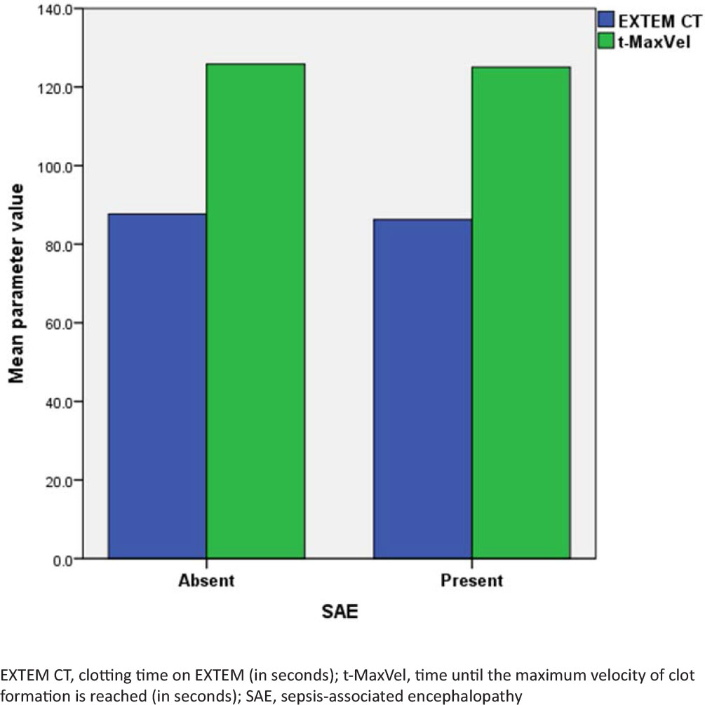
Fig. 1.
Comparison of coagulation initiation between groups.
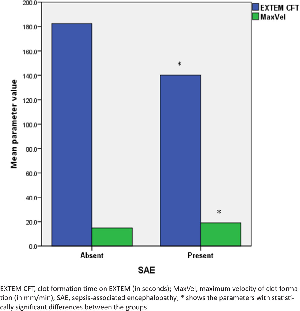
Fig. 2.
Comparison of coagulation propagation between groups.
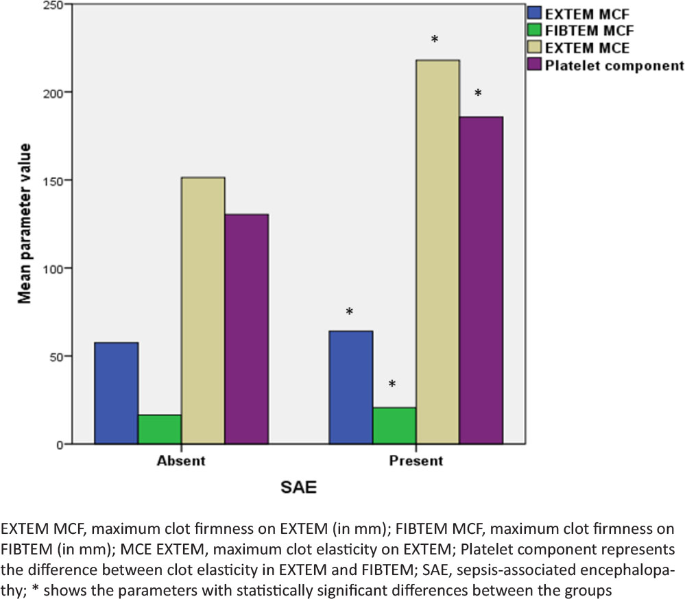
Fig. 3.
Comparison of clot strength between groups.
Table 4.
Multivariate logistic regression model using fibrinogen levels and platelet contribution to clot strength.
| Parameter | Adjusted OR | 95% CI | P value |
|---|---|---|---|
| Fibrinogen level | 1.000 | 0.997; 1.002 | 0.403 |
| Platelet component | 1.007 | 1.001; 1.013 | 0.02* |