
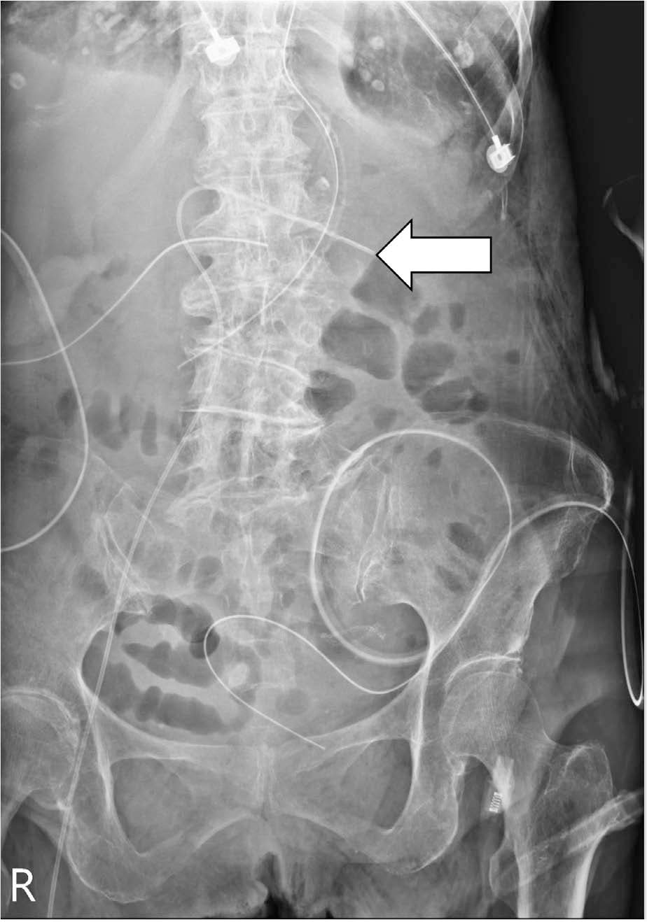
Fig. 1.
Postprocedural abdominal X-ray showing misplacement of the catheter tip in the contralateral renal vein (arrow).
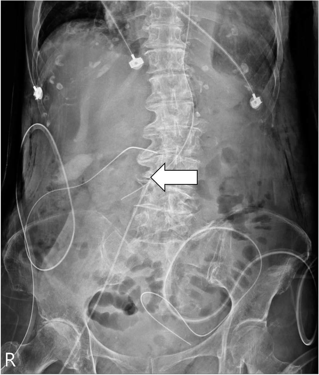
Fig. 2.
Relocation of the catheter tip within the middle segment of the inferior vena cava after pulling out by 8 cm (arrow).
Table 1.
Previously reported malposition cases with SFV PICCs
| First author (year) | Number of patients | Number of malposition cases (%) | Age/Sex | Malposition place | Subsequent management | Result of interest |
|---|---|---|---|---|---|---|
| Wan (2018) | 221 | 0 (0) | n/a | n/a | n/a | Significant lower malposition rate in patients with SFV PICCs (0%) compared to upper limb PICCs (2.15%). |
| Zhao (2019) | 121 | 2 (1.65) | 89/f | Contralateral common iliac vein | No adjustment or re-puncture based on the decision from the doctor and the patient’s guardian. | Significant lower malposition rate in patients with SFV PICCs (1.65%) compared to upper limb PICCs (6.02%). |
| 73/m | Folded at ipsilateral common iliac vein | Pull out 4 cm according to the X-ray findings. | ||||
| Elli (2022) | 142 | 4 (2.82) | n/a | n/a | Immediate replacement | n/a |