
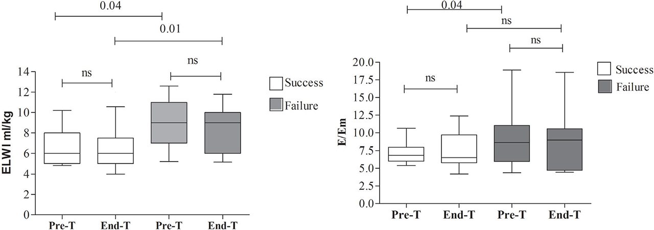
Fig. 1.
Extravascular lung water index (EVLWI) and E/Tissue Doppler Imaging early diastolic velocity (E/Em) in patients undergoing a spontaneous breathing trial before (Pre SBT) and following (End SBT) the trial, according to trial outcome (Median values, inter-quartile range and 10–90th percentiles are presented).
Table 1.
Baseline characteristics of participants according to spontaneous breathing trial outcome.
| Weaning success (N=14) | Weaning failure | P-value | ||
|---|---|---|---|---|
| EWF (<2h) (N=5) | LWF (2-48h) (N=6) | |||
| Age, years | 58.31 (4.47) | 63.25 (2.95) | 67.83 (6.84) | 0.45 |
| Gender (male) | 11 (78.5) | 3 (60) | 4 (66.7) | 0.69 |
| Medical /Surgical critical state | 3 (21.5) / 11 (78.5) | 2 (40) / 3 (60) | 4 (66.7) /2 (33.3) | 0.08 |
| Sepsis | 3 | 2 | 1 | |
| Intoxication | 0 | 0 | 1 | |
| Pyelonephritis | 0 | 0 | 1 | |
| Status epilepticus | 0 | 0 | 1 | |
| Peritonitis | 0 | 0 | 1 | |
| Pancreatitis | 1 | 0 | 0 | |
| Neurosurgical | 5 | 2 | 1 | |
| Multiple trauma | 2 | 0 | 0 | |
| Cancer | 1 | 1 | 0 | |
| Hemorrhage | 2 | 0 | 0 | |
| Arterial Hypertension | 9 (64.3) | 5 (100) | 5 (83.3) | 0.24 |
| COPD | 1 (7) | 1 (20) | 0 | 0.46 |
| Diabetes mellitus | 3 (21) | 1 (20) | 0 | 0.47 |
| APACHE II (on admission) | 14.9 (2.66) | 18 (4.37) | 8.33 (5.84) | 0.36 |
| SOFA (on admission) | 8.54 (1.64) | 7.25 (1.65) | 9 (1.46) | 0.86 |
| Fluid balance, mL/day | 2676 (630) | 472 (1667) | 2513 (1564) | 0.35 |
| Renal dysfunction | 3 (21) | 2 (40) | 2 (33.3) | 0.69 |
| T-piece trial | ||||
| ICU day of SBT | 22.79 (6.06) | 16 (6) | 17.33 (2.65) | 0.72 |
| Orotracheal/Tracheostomy tube | 11(78) / 3(21) | 3(60) / 2(40) | 4 (67) / 2(33) | 0.69 |
| SOFA | 5.3 (0.9) | 3.2 (1.1) | 3.8 (0.7) | 0.38 |
| PaO2/FIO2 ratio | 370 (31) | 281 (78) | 392 (36) | 0.16 |
| PEEP, cmH2O | 6.3 (0.3) | 7 (0.4) | 6.7 (0.4) | 0.52 |
| f, breaths/min | 19.7 (1.8) | 24.2 (1.4) | 21.1 (1.3) | 0.24 |
| VT, L | 0.47 (0.014) | 0.46 (0.017) | 0.51 (0.026) | 0.29 |
| f /VT (RSBI) | 41.7 (3.9) | 52.9 (4.7) | 42.3 (3.7) | 0.20 |
| PFP, cmH2O*breaths/min | 264 (201) | 316 (27) | 280 (27) | 0.52 |
| MIP, cmH2O | 23.6 (1.57) | 23.2 (1.83) | 25.71 (1.83) | 0.63 |
| BNP, pg/mL | 412 (157) | 761 (728) | 455 (125) | 0.70 |
| Albumin, mg/dL | 2.7 (0.1) | 3 (0.2) | 2.8 (0.1) | 0.32 |
* Spontaneous Breathing Trial-induced significant changes within subgroups (pre-trial vs, end-trial measurements), examined by paired t-test analysis
EWF= early weaning (T-trial) failure; LWF= late weaning failure; ANOVA=analysis of variance; COPD= chronic obstructive pulmonary disease; APACHE II=Acute Physiology and Chronic Health Evaluation Score II; SOFA score=Sequential Organ Failure Assessment score; PEEP= positive end-expiratory pressure; f= respiratory frequency; VT= tidal volume; RSBI= rapid shallow breathing index; PFP (cmH2O*breaths/min) = pressure frequency product (respiratory frequency x level of pressure support); renal dysfunction= creatinine level≥2mg/dL at weaning; MIP= maximal inspiratory pressure; MIP percentage = percentage of measured MIP in relation to predicted MIP according to age and sex; BNP=B-type natriuretic peptide; Renal ICU= Intensive Care Unit.
Table 2.
PiCCO hemodynamics according to spontaneous breathing trial outcome before (Pre-T) and following (End-T) the trial.
| Weaning success (N=14) | Weaning failure (N=11) | P-value | ||
|---|---|---|---|---|
| MAP, mmHg | Pre-T | 86.5 (2.6) | 95.2 (2.6) | 0.02 |
| End-T | 91.1 (3.2) | 100.4 (6.6) | NS | |
| HR, beats/min | Pre-T | 92.1 (4.8) | 82.82 (5.2) | NS |
| End-T | 95.9 (4.4) | 94.0 (57.6)* | NS | |
| RPP, beats*min−1*mmHg | Pre-T | 7974 (502) | 7985 (603) | NS |
| End-T | 8786 (550) | 9648 (1072) | NS | |
| ScvO2, % | Pre-T | 76.6 (1.7) | 74.5 (3) | NS |
| End-T | 79 (1.5) | 72.6 (2.5) | 0.03 | |
| O2ER, % | Pre-T | 22.8 (1.6) | 24.4 (2.9) | NS |
| End-T | 19.3 (1.5)* | 24.3 (2.2) | NS | |
| CVP, mmHg | Pre-T | 5.43 (1) | 11.27 (1.9) | 0.01 |
| End-T | 4.86 (0.9) | 10.82 (1.6) | 0.01 | |
| EVLW, mL | Pre-T | 430 (20.4) | 603 (56) | 0.01 |
| End-T | 440 (30) | 566.4 (43) | 0.01 | |
| ELWI, mL/kg | Pre-T | 6.79 (0.7) | 9 (0.7) | 0.04 |
| End-T | 6.21 (0.6) | 8.54 (0.7) | 0.01 | |
| CI, L/min/m2 | Pre-T | 3.82 (0.3) | 3.63 (0.3) | NS |
| End-T | 4.22 (0.3)* | 4.25 (0.4)* | NS | |
| SVI, mL/m2 | Pre-T | 45.3 (3.4) | 43.9 (4.8) | NS |
| End-T | 48.1 (3.3) | 47 (4.3) | NS | |
| GEDI, mL/m2 | Pre-T | 659 (19) | 846 (75) | 0.013 |
| End-T | 691 (31) | 861 (80) | 0.04 | |
| PBV, mL | Pre-T | 349.3(12.4) | 452 (82.6) | NS |
| End-T | 367.7(15.9) | 410 (45.6) | NS | |
| SVRI, cm.d.sec−5m2 | Pre-T | 1833 (108) | 2122 (199) | NS |
| End-T | 1755 (101) | 1924 (222) | NS | |
| dPmx, mmHg/min | Pre-T | 1357 (118) | 1070 (130) | NS |
| End-T | 1266 (154) | 1146 (113) | NS | |
| PVP | Pre-T | 1.28 (0.1) | 1.65 (0.2) | 0.028 |
| End-T | 1.22 (0.1) | 1.5 (0.2) | NS | |
| CFI, L/min | Pre-T | 5.79 (0.4) | 4.58 (0.6) | NS |
| End-T | 6.16 (0.4) | 5.25 (0.6) | NS | |
| GEF, % | Pre-T | 52.2 (3.8)* | 42.36 (4)* | NS |
| End-T | 26.7 (0.9)* | 23.94 (3)* | NS |
Data represent mean±SE otherwise is indicated. P-values represents significant differences (P≤0.05) between weaning success and weaning failure subgroups.
* Significant differences (P≤0.05) between Pre-T and End-T values within subgroups of weaning outcome.
MAP= mean arterial pressure; Pre-T=pre-trial; End-T= end-trial; HR= heart rate; ScvO2= caval oxygen saturation; O2ER=oxygen extraction ratio; CVP= central venous pressure; EVLW= extravascular lung water; ELWI= extra-vascular lung water, indexed to predicted body weight; CI= cardiac index; SV= stroke volume index; SVI= stroke volume, indexed to body surface area; GEDV= global end-diastolic volume; GEDI= global end-diastolic volume, indexed to body surface area; PBV= pulmonary blood volume; SVRI= systemic vascular resistance index; dPmx= maximum pressure increase in the aorta (ΔPmax/Δt); PVP= pulmonary vascular permeability; PVPI= pulmonary vascular permeability, indexed to body surface area; SVV= stroke volume variation; CFI= cardiac function index; GEF= global ejection fraction RPP= Rate-pressure product.
Table 3.
Echocardiographic parameters in T-piece weaning trial in respect to T-trial and weaning outcomes.
| SBT-outcome | Weaning Outcome | ||||||
|---|---|---|---|---|---|---|---|
| Success, n=20 | Failure, n=5 | P value | Success, n=14 | Failure, n=11 | P value | ||
| Right Ventricular Echocardiography | |||||||
| RVFAC, % | Pre-T | 44.2 (2.1) | 33.32 (16) | - | 46.26 (2.2) | 37.32(6.5) | - |
| RV/LV EDA | Pre-T | 0.58 (0.04) | 0.38 (0.03) | 0.02 | 0.54 (0.04) | 0.54(0.06) | - |
| RV Sm, cm/sec | Pre-T | 16.61 (0.8) | 13.25 (0.9) | - | 17.08 (0.96) | 14.9(0.9) | - |
| End-T | 16.39 (0.9) | 14.25 (0.7) | - | 16.93 (1.24) | 14.9(0.7) | - | |
| RV Em, cm/sec | Pre-T | 13.49(1.33) | 13.38 (2) | - | 14.09 (1.58) | 12.3 (1.2) | - |
| End-T | 12.33(0.93) | 12.37 (0.8) | - | 13.39 (1.05) | 12.3(1.2) | - | |
| RV Am, cm/sec | Pre-T | 17.37 (1.5) | 18.97 (2.3) | - | 16.8 (1.49) | 11 (1) | - |
| End-T | 18.27 (1.1) | 22.3 (2.43) | - | 18.56 (1.23) | 19.1 (1.7) | - | |
| RV IVA, m/sec2 | Pre-T | 5.22 (0.59) | 2.97 (0.71) | - | 5.76 (0.66) | 3.7 (0.5) | 0.021 |
| End-T | 4.44 (0.65) | 3.21 (0.12) | - | 4.93 (0.83) | 2.9 (0.15) | 0.05 | |
| RV Tei index | Pre-T | 0.66 (0.08) | 0.65 (0.15) | - | 0.67 (0.1) | 0.66(0.1) | - |
| End-T | 0.79 (1.13) | 0.6 (0.05) | - | 0.83 (0.18) | 0.64(0.1) | - | |
| Left Ventricular Echocardiography | |||||||
| LVEF, % | Pre-T | 62.63 (2.1) | 58.22 (8.1) | - | 63.14 (2.9) | 60 (3.7) | - |
| LV mass, g/m2(Penn) | Pre-T | 270 (27) | 268 (58) | - | 267.9 (39.5) | - | |
| IVSWT, mm | Pre-T | 10.8 (0.7) | 9.4 (0.6) | - | 11 (1) | 10 (0.4) | - |
| LVPWT, mm | Pre-T | 9.9 (0.5) | 8.7 (0.3) | - | 10 (0.7) | 9.5 (0.3) | - |
| LVIDd, mm | Pre-T | 45.5 (1) | 49.1 (5) | - | 44.6 (1.4) | 48.2 (1.9) | - |
| LVIDs, cm | Pre-T | 30 (0.7) | 34.3 (5.3) | 28.7 (0.6) | 33.5 (0.2) | 0.024 | |
| E, cm/sec | Pre-T | 72.28 (4.9) | 83.3 (8.6) | - | 67.18 (3.14) | 83.8 (8.3) | 0.05 |
| End-T | 80.71(5.7) * | 95.1 (8.9 * | - | 76.14(3) * | 93.1(10) * | - | |
| A, cm/sec | Pre-T | 76.98 (4.8) | 88.7 (7.4) | 70.31 (4.36) | 90.2 (6.2) | 0.014 | |
| End-T | 81.63 (5.2) | 97.6 (7.8) | - | 78.51 (4.76) | 93.6 (7.3) | - | |
| DTE, msec | Pre-T | 212.4(14.4) | 169.5 (27) | - | 198.6 (11.7) | 211.6(22) | - |
| End-T | 190.9 (12) | 154.8 (24) | - | 179.82 (11) | 183(19.5) | - | |
| E/A | Pre-T | 0.96 (0.05) | 0.97 (0.13) | - | 0.96 (0.05) | 0.95(0.1) | - |
| End-T | 1 (0.05) | 1.02 (0.17) | - | 0.99 (0.04) | 1(0.1) | - | |
| LV Sm, cm/sec | Pre-T | 11.71 (0.6) | 10.27 (1.6) | - | 11.42 (0.8) | 10.3(0.8) | - |
| End-T | 11.94 (0.8) | 11.95 (1.4) | - | 12.98 (1) | 10.7(0.8) | - | |
| LV Em, cm/sec | Pre-T | 10.07 (0.7) | 9.16 (0.9) | - | 10.05 (0.6) | 9.5 (1) | - |
| End-T | 11.62(0.9) * | 11.1 (1.4) * | - | 12.1 (1) * | 11 (1.3) * | - | |
| LV Am, cm/sec | Pre-T | 9.7 (0.4) | 12 (0.9) | 0.003 | 10.08 (0.43) | 10.6(0.8) | - |
| End-T | 11.1 (0.8) | 13.24 (1.4) | - | 11.22 (1) | 11.9 (1) | - | |
| E/Em | Pre-T | 7.87 (0.9) | 9.56 (1.5) | 6.87 (0.35) | 9.8 (1.6) | 0.05 | |
| End-T | 7.85 (0.1) | 9.08 (1.32) | - | 6.73 (0.6) | 9.72 (1.7) | - | |
| LV IVA, m/sec2 | Pre-T | 3.58 (0.39) | 2.69 (0.37) | - | 3.8 (0.47) | 2.6 (0.2) | 0.046 |
| End-T | 3.37 (0.4) | 2.67 (0.47) | - | 3.8 (0.47) | 2.5 (0.36) | 0.05 | |
| LV Tei index | Pre-T | 0.64 (0.06) | 0.8 (0.15) | - | 0.62 (0.09) | 0.72(0.1) | - |
| End-T | 0.71 (0.11) | 0.75 (0.2) | - | 0.73 (0.17) | 0.7 (0.1) | - | |
* Spontaneous Breatning Trial-induced significant changes within subgroups, examined by paired t-test analysis.
RV=right ventricular; RVFAC= right ventricular fractional area change; LV= left ventricular; EDA= end-diastolic area; RV Sm, Em, Am = Tissue Doppler Imaging (TDI)-derived peak systolic, early and late diastolic velocity, respectively (measured at the lateral border of tricuspid annulus); Pre-T=pre-trial; End-T= end-trial; IVA= isovolumic acceleration velocity; LVEF= Left ventricular ejection fraction; IVSWT= inter-ventricular septum wall thickness; LVPWT= left ventricular posterior wall thickness; LVIDd= left ventricular internal diameter at end-diastole; LVIDs=left ventricular internal diameter at end-systole; E and A= pulsed wave Doppler early and late transmitral left ventricular filling wave velocity, respectively; DTE= E–wave deceleration time; LV Sm, Em, Am = Tissue Doppler Imaging (TDI)-derived peak systolic, early and late diastolic velocity, respectively (measured at the lateral border of mitral annulus).
Table 4.
Univariate and independent determinants of extravascular lung water index (pre spontaneous breathing trial values)
| Univariate linear regression analysis | Multivariate linear regression analysis | |||||
|---|---|---|---|---|---|---|
| r | R2 | P value | b | B (95%CI) | P value | |
| GEF | −0.481 | 0.231 | 0.03 | −0.013 | −0.003 (−0.024 – 0.019) | NS |
| LV Sm | −0.487 | 0.237 | 0.01 | 0.004 | 0.004 (−0.131 – 0.139) | NS |
| LV E/Em | 0.404 | 0.163 | 0.04 | 0.203 | 0.133 (0.063 – 0.202) | 0.01 |
| CVP | 0.431 | 0.185 | 0.03 | −0.041 | −0.018 (−0.066 – 0.031) | NS |
[i] ELWI= extravascular lung water index; r=Pearson’s correlation coefficient; R2= coefficient of determination; B, b= unstandardized and standardized beta coefficients, respectively; CEF=global ejection fraction; LV=left ventricular; Sm, Em= peak systolic and early diastolic tissue-Doppler myocardial velocities at the lateral border of the mitral annulus, respectively; E=E-wave mitral inflow pulsed-wave Doppler velocity; CVP=central venous pressure
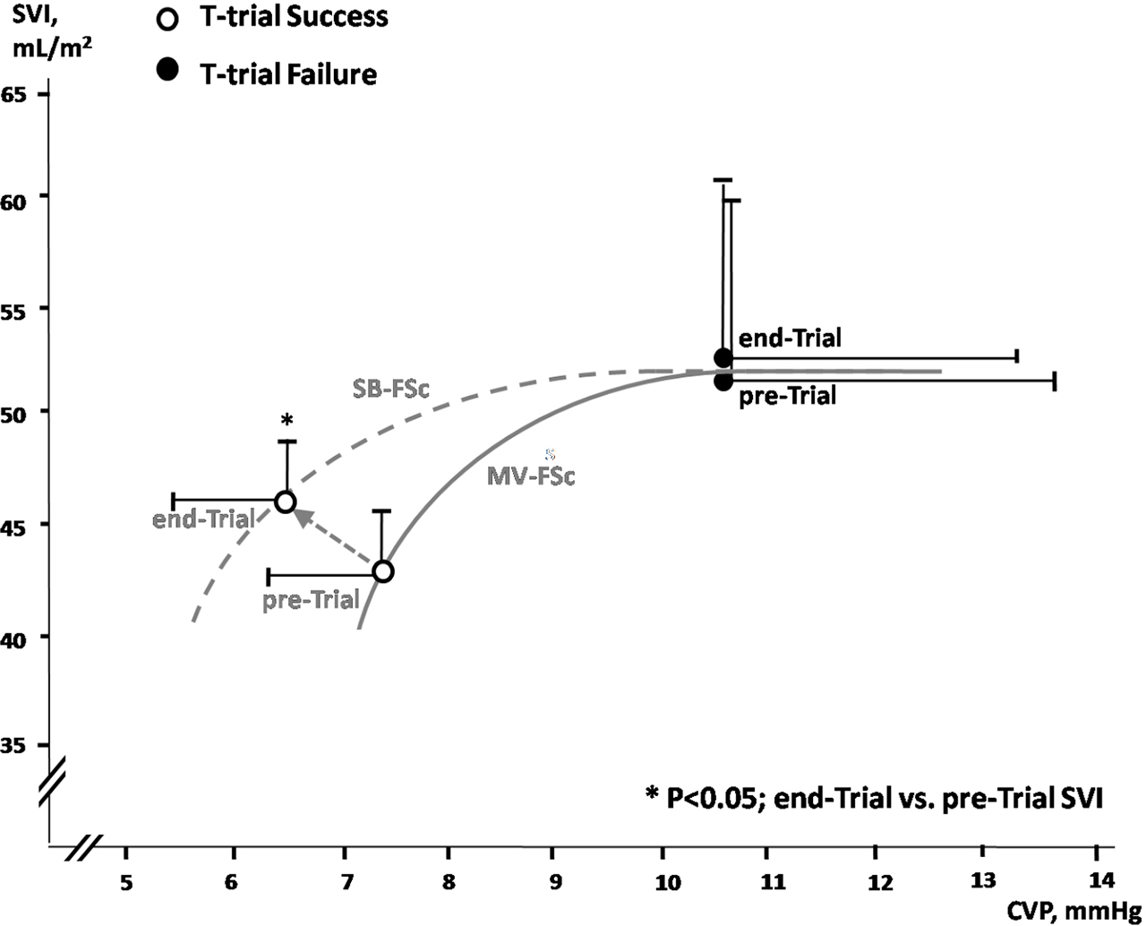
Fig. 2.
Stroke Volume Index (SVI) against central venous pressure (CVP) plot and Frank Starling curve representations in participants during spontaneous breathing and mechanical ventilation. Open circles represent successful weaning, closed circles represent weaning failure; SB-FSc=Frank-Starling’s curve at spontaneous breathing; MV-FSc= Frank-Starling’s curve during mechanical ventilation.