
Table 1.
Urine analysis and immunology tests performed on the third day following admission of the patients to the pediatric nephrology department
| Analysis | Case 1 | Case 2 |
|---|---|---|
| Urinalysis | ||
| Erythrocytes | 300/μL | 300/μL |
| Urine sediment | ||
| Erythrocytes | 11–20/field | 21–50/field |
| 80% dysmorphic red blood cells, 20% isomorphic red blood cells /field | 95% dysmorphic red blood cells, 5% isomorphic red blood cells /field | |
| Hematic casts | ||
| Leukocytes | 0–5/field | 0–5/field |
| Proteins | negative | 1 g/L |
| C3 | 0,33 g/L | 0,19 g/L |
| C4 | 0,104 g/L | 0.123 g/L |
| C3 after 6 weeks | 1,48 g/L | 1,17 g/L |
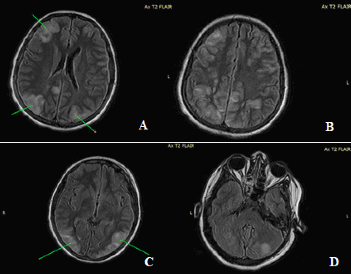
Fig. 1.
Multiple infra and supra centimeter lesions in hypersignal T2 and FLAIR diffusely arranged bihemispheric cortico-subcortical in (A, B) frontal and parietal lobes, (C) occipital lobe and (D) bihemispheric cerebellar
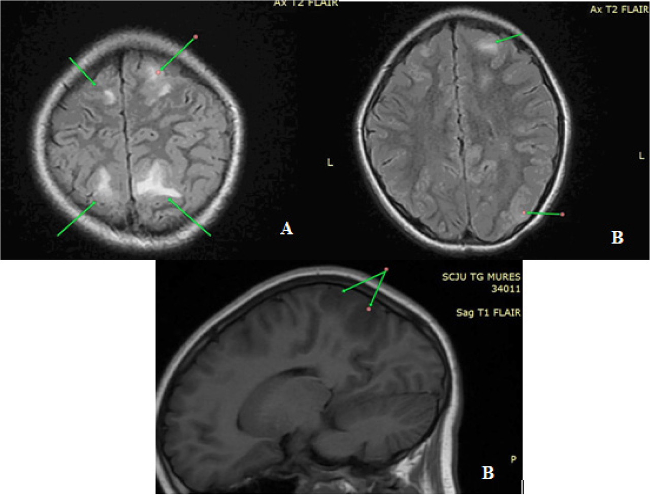
Fig. 2.
An inflammatory cerebral lesion showing hypersignal on T2 and FLAIR sequences, located at the level of the bilateral frontal lobe and post-central gyrus (A), respectively on the left parietal side (B)