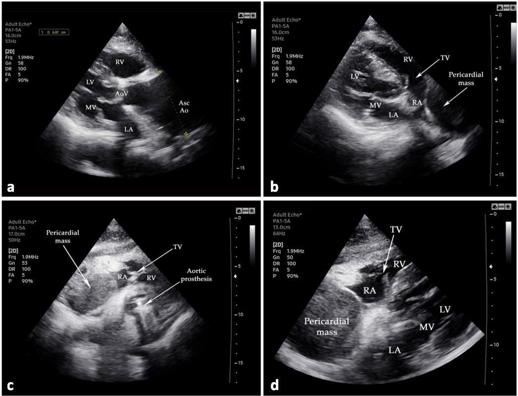
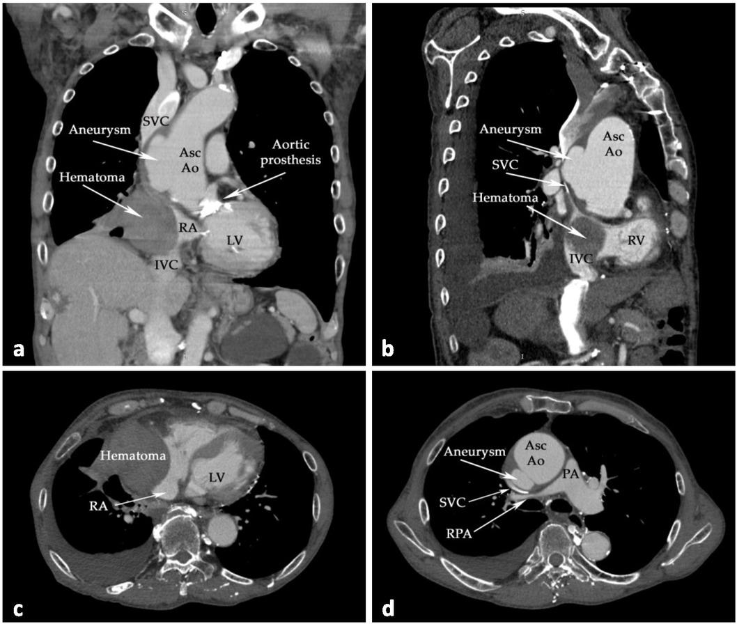
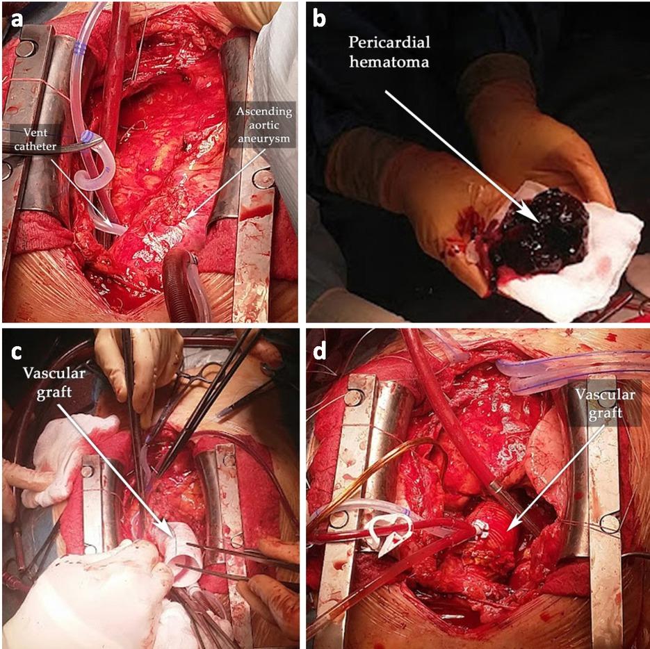
Have a personal or library account? Click to login
Right Heart Failure as an Atypical Presentation of Chronic Type A Aortic Dissection - Multimodality Imaging for Accurate Diagnosis and Treatment. A case report and mini-review of literature Cover

Right Heart Failure as an Atypical Presentation of Chronic Type A Aortic Dissection - Multimodality Imaging for Accurate Diagnosis and Treatment. A case report and mini-review of literature

Open Access
|Aug 2022

Figures & Tables

Fig. 1

Transthoracic echocardiography examination: (a) the clear presence of the ascending aortic aneurysm (6.87 cm); (b–d) massive pericardial tumoral mass compressing the right atrium. Abbreviations: LA—left atrium; LV—left ventricle; MV—mitral valve; RA—right atrium; RV—right ventricle; SVC—superior vena cava; TV—tricuspid valve; AoV — aortic valve; AscAo — ascending aorta.

Fig. 2

Emblematic thoraco-abdominal CT scans. In the coronal and sagittal scans ((a) and (b), respectively), the presence of an ascending aortic aneurysm, hematoma, and compression on RA, SVC, and IVC can be observed. (c) Compression of RA; (d) compression of SVC and RPA (axial CCTA scans). Abbreviations: AscAo—ascending aorta; IVC—inferior vena cava; LV—left ventricle; PA—main pulmonary artery; RA—right atrium; RPA—right pulmonary artery; SVC—superior vena cava.

Fig. 3

Intraoperative images. (a) Post-sternotomy, the ascending aortic aneurysm was revealed; (b) the chronic pericardial hematoma; (c) the ascending aorta replaced with a 30 mm-diameter vascular graft; (d) the final result.
DOI: https://doi.org/10.2478/jccm-2022-0016 | Journal eISSN: 2393-1817 | Journal ISSN: 2393-1809
Language: English
Page range: 204 - 213
Submitted on: Apr 9, 2022
Accepted on: Jun 12, 2022
Published on: Aug 12, 2022
Published by: University of Medicine, Pharmacy, Science and Technology of Targu Mures
In partnership with: Paradigm Publishing Services
Publication frequency: 4 issues per year

© 2022 Ioan Tilea, Robert Adrian Dumbrava, Alexandra Mihaela Ratiu, Marius Mihai Harpa, Cosmin Marian Banceu, Dorina Nastasia Petra, Horatiu Suciu, published by University of Medicine, Pharmacy, Science and Technology of Targu Mures
This work is licensed under the Creative Commons Attribution 4.0 License.