
Table 1
The primary stroke timelines in the patient’s case history
| The main events in the patient’s stroke history | Time |
|---|---|
| the appearance of the first symptoms: cervical pain, headache, dizziness | 19:00 |
| the patient was last seen without neurological deficit | 23:30 |
| the patient was found unconscious with difficult respiration | 24:00 |
| presentation to the emergency unit | 01:48 |
| emergency laboratory workup | 01:55 |
| cerebral CT scan | 01:56 |
| cervico-cerebral angio CT scan | 02:24 |
| groin picture | 03:47 |
| recanalization | 04:50 |
| the patient was transferred to the intensive care unit | 05:50 |
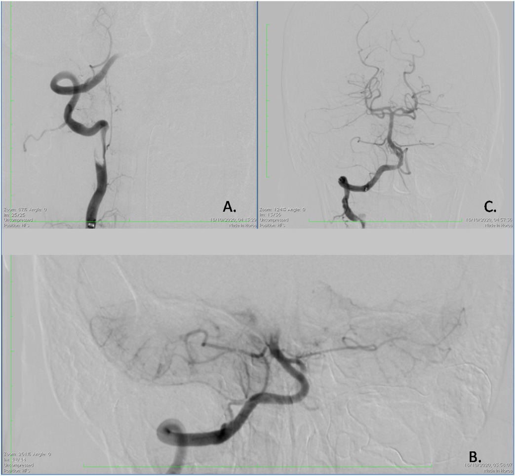
Fig. 1
Digital subtraction angiography. A. Long, eccentric, irregular stenosis (string sign) at the distal V2 segment of the right vertebral artery; B. Basilar artery occlusion 8 mm from the origin; C. Complete recanalization of the basilar artery after thrombectomy
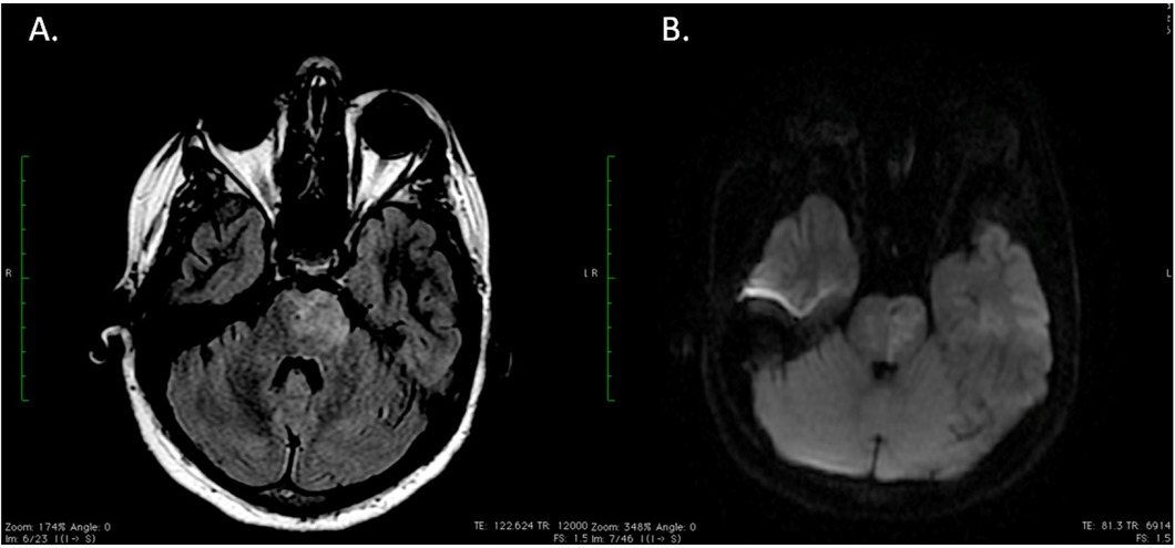
Fig. 2
A. Axial FLAIR weighted; B. Axial Diffusion-weighted MRI images showing bilateral, predominantly left-sided pontine infarction.