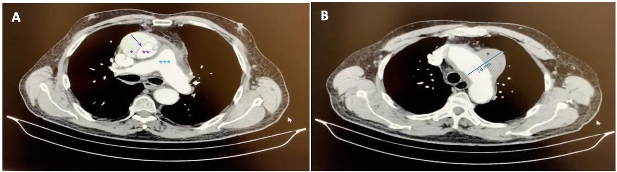
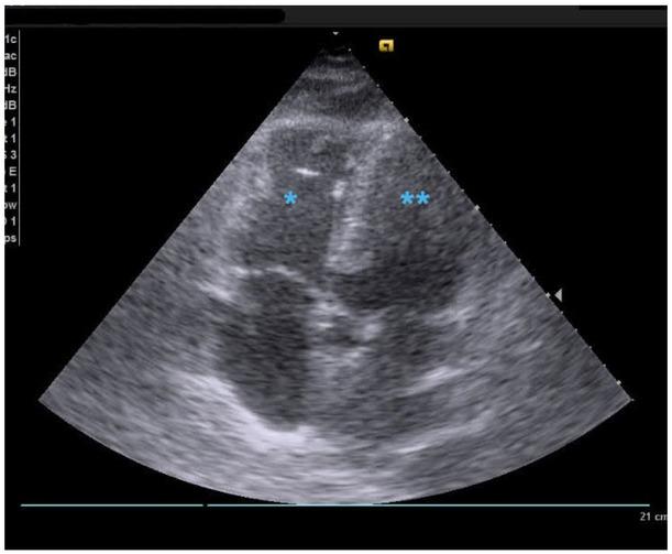
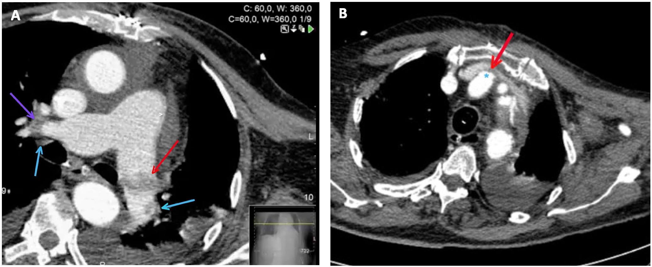
Have a personal or library account? Click to login
An Incident of a Massive Pulmonary Embolism following Acute Aortic Dissection. A Case Report Cover

An Incident of a Massive Pulmonary Embolism following Acute Aortic Dissection. A Case Report

Open Access
|Jan 2021

Figures & Tables

Fig. 1

A. Preoperative thoracic computed tomographic angiography: acute Stanford type A aortic dissection with ascending intimal flap (arrow). The main pulmonary artery(***) and the left and right branches were normally enhanced with contrast medium. (*true lumen aorta; **false lumen). B. Preoperative thoracic computed tomographic angiography: aortic arch saccular aneurysm (*).

Fig. 2

Transthoracic echocardiography apical five-chamber view: right ventricular enlargement (*) and left ventricle (**).

Fig. 3

A. Contrast-enhanced thoracic CT showing extensive, bilateral thromboembolism of the right (purple arrow) and left pulmonary artery (red arrow) branches and its lobar branches (blue arrows). B. Periprosthetic hematoma from the innominate artery with a pseudoaneurysm (*) conditioning a slight impression of the brachiocephalic venous trunk filiform path (red arrow).
DOI: https://doi.org/10.2478/jccm-2021-0001 | Journal eISSN: 2393-1817 | Journal ISSN: 2393-1809
Language: English
Page range: 67 - 72
Submitted on: Sep 14, 2020
|
Accepted on: Dec 29, 2020
|
Published on: Jan 29, 2021
In partnership with: Paradigm Publishing Services
Publication frequency: 4 issues per year

© 2021 Sofia Fernandes, Mariana Rodrigues, Catarina Barreiros, Hugo Côrte-Real, Ricardo Ferreira, Ângelo Nobre, published by University of Medicine, Pharmacy, Science and Technology of Targu Mures
This work is licensed under the Creative Commons Attribution 4.0 License.