
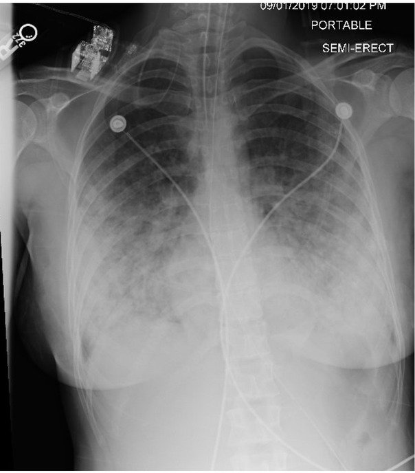
Fig. 1
Chest radiograph (CXR) demonstrating bilateral peripheral and central opacities with a basilar predominance
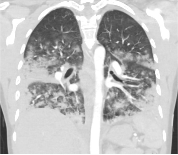
Fig. 2
Chest computerized tomography (CT) demonstrating diffuse, broncho-centric bilateral ground glass opacities with coalescence to consolidation, largely in the lung bases
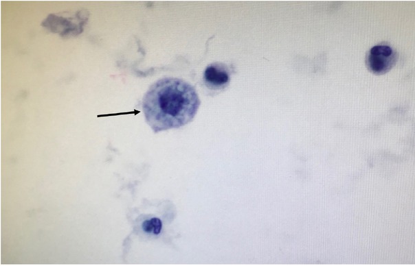
Fig. 3
Lung cytopathology, demonstrating rare foamy macrophages (black arrow).
Table 1
Diagnostic criteria for acute eosinophilic pneumonia.
| Clinical diagnostic criteria for acute eosinophilic pneumonia |
|---|
| Febrile illness less than four weeks in duration |
| Hypoxemic respiratory failure |
| Diffuse pulmonary opacities on chest radiograph |
| Bronchoalveolar lavage (BAL) with cell count demonstrating >25% eosinophilia |
| Absence of other known causes for pulmonary eosinophilia |
Table 2
Differential diagnosis considerations for acute eosinophilic pneumonia.
| Differential diagnosis considerations |
|---|
| Eosinophilic granulomatosis with polyangiitis |
| Pulmonary fungal infections |
| Pulmonary parasitic infections |
| Medication effects |
| Pulmonary infiltration of eosinophils secondary to overwhelming peripheral eosinophilia |