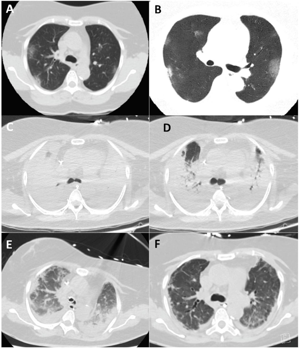
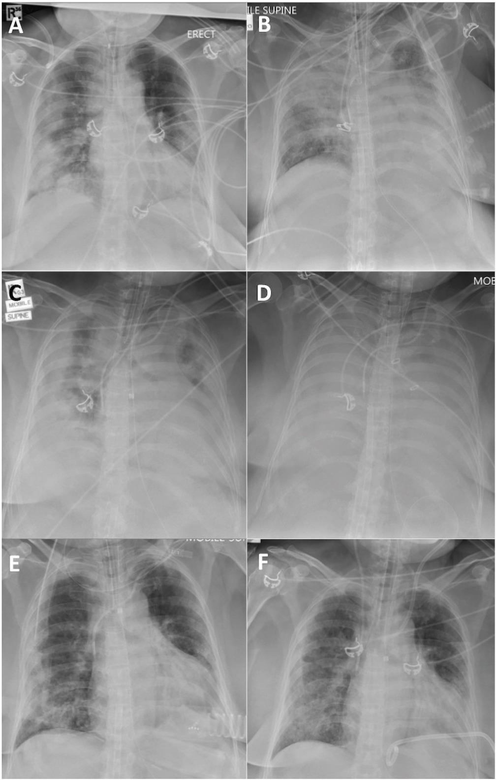
Have a personal or library account? Click to login
The Prolonged Use of VV ECMO Support in COVID-19: A Case Report Cover

Figures & Tables

Fig.1

CT Chest Axial Imaging demonstrating progression and resolution of lung infiltration. Image A, B - Admission to hospital; C,D - Imaging performed on PEEP of 5 cmH2O and 40 cmH2O respectively;D - on day 13 of ECMO; E- Day 29 of ECMO; F - 13 days post decannulation.

Fig.2

Plain Chest radiographs demonstrating progression and resolution of lung infiltration. A - post intubation; B - initiation of ECMO; C - Day 13 of ECMO; D - Day 20 of ECMO after initiation of proning; E - Day 34 of ECMO; F - 13 days post ECMO decannulation.
DOI: https://doi.org/10.2478/jccm-2020-0034 | Journal eISSN: 2393-1817 | Journal ISSN: 2393-1809
Language: English
Page range: 224 - 230
Submitted on: Aug 2, 2020
Accepted on: Sep 29, 2020
Published on: Nov 7, 2020
Published by: University of Medicine, Pharmacy, Science and Technology of Targu Mures
In partnership with: Paradigm Publishing Services
Publication frequency: 4 issues per year

© 2020 Amelia Lucy Fitzgerald, Hemal Hitesh Vachharajani, Benjamin Paul Davidson, Natalie Joanne Kruit, Adam Trevor Eslick, published by University of Medicine, Pharmacy, Science and Technology of Targu Mures
This work is licensed under the Creative Commons Attribution-NonCommercial-NoDerivatives 4.0 License.