
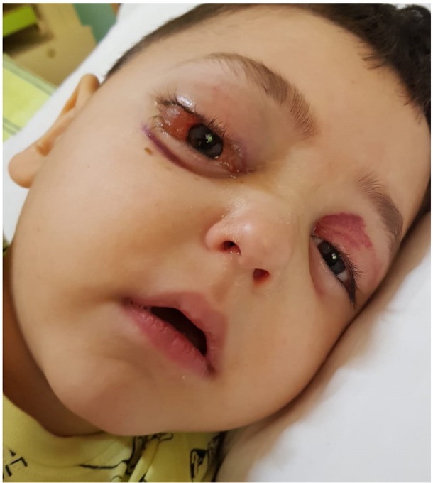
Fig. 1
Proptosis of the right eye in the first 24 hours after admission to the Paediatric Clinic
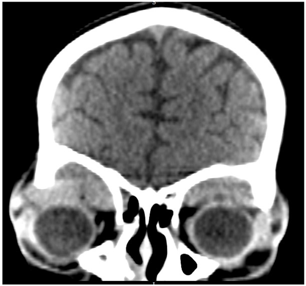
Fig. 2
The non-contrast-enhanced cranial/cerebral CT of the head with coronal sections at the level of the orbits, shows two well-defined, soft-tissue density lesions in the supero-lateral aspect of the extraconal regions bilaterally, more pronounced on the right side, where downward displacement of the ocular globe is visible
Table 1
Routine tests results performed on Day 1 post-admission to Paediatrics Clinic
| CRP | Fg | ESR | LDH | AST | ALT | Creatinine | Urea |
|---|---|---|---|---|---|---|---|
| 41.66 mg/l | 497 mg/dl | 93 mm/h | 601 U/l | 21 U/l | 8 U/l | 0,69 mg/dl | 26,1 mg/dl |
Table 2
Bone marrow immunophenotyping
| CD34 | HLA-DR | CD13 | CD33 | CD15 | CD117 | CD19 | CD3 | CD7 | CD10 | CD22 | CD11 | CD14 | CD64 |
|---|---|---|---|---|---|---|---|---|---|---|---|---|---|
| 80% | 80% | 78% | 75% | 70% | 60% | 62% | negative | negative | negative | negative | negative | negative | negative |
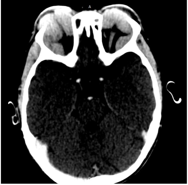
Fig. 3
Contrast-enhanced cranial CT axial view of the head and orbit, following intravenous iodinated contrast media administration. The lesions show mild, homogenous enhancement.
Table 3
Supportive care of the induction phase.
| Drug name | Strength | Dosage | Route of administration | Dates of administration |
|---|---|---|---|---|
| human immune globulin | 2,5g/25ml | 1 fl/day | intravenously | Day 13 |
| (CSL Behring GmbH, Marburg, Germany) | ||||
| red blood cell | 10ml/kg | intravenously | Day 9,21 | |
| pooled platelets | 1unit/10kg | intravenously | Day 7-16, 21-25 | |
| trimethoprim-sulfamethoxazole | (40mg/200mg)/5ml | 2x10ml/day | orally | Day 24-26 |
| (E.I.P.I.Co., Tenth of Ramadan City, Egypt) | ||||
| fluconazole | 2mg/ml | 100 mg once | intravenously | Day 17-27 |
| (Fareva Amboise, Pocé-Sur-Cisse, France) | daily | |||
| granisetron | 1mg/ml | 1mg twice a | Intravenously | Day 4-11 |
| (Fresenius Kabi Austria GmbH, Graz, Austria) | day | |||