
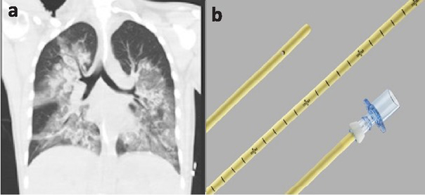
Fig.1
a. Admission chest CT (coronal view) showing multilobar consolidation and areas of ground-glass opacification in all five lobes. b. Cook® Airway Exchange Catheter & Rapi-Fit® adapter with 15-mm connector for oxygenation during exchange procedures.
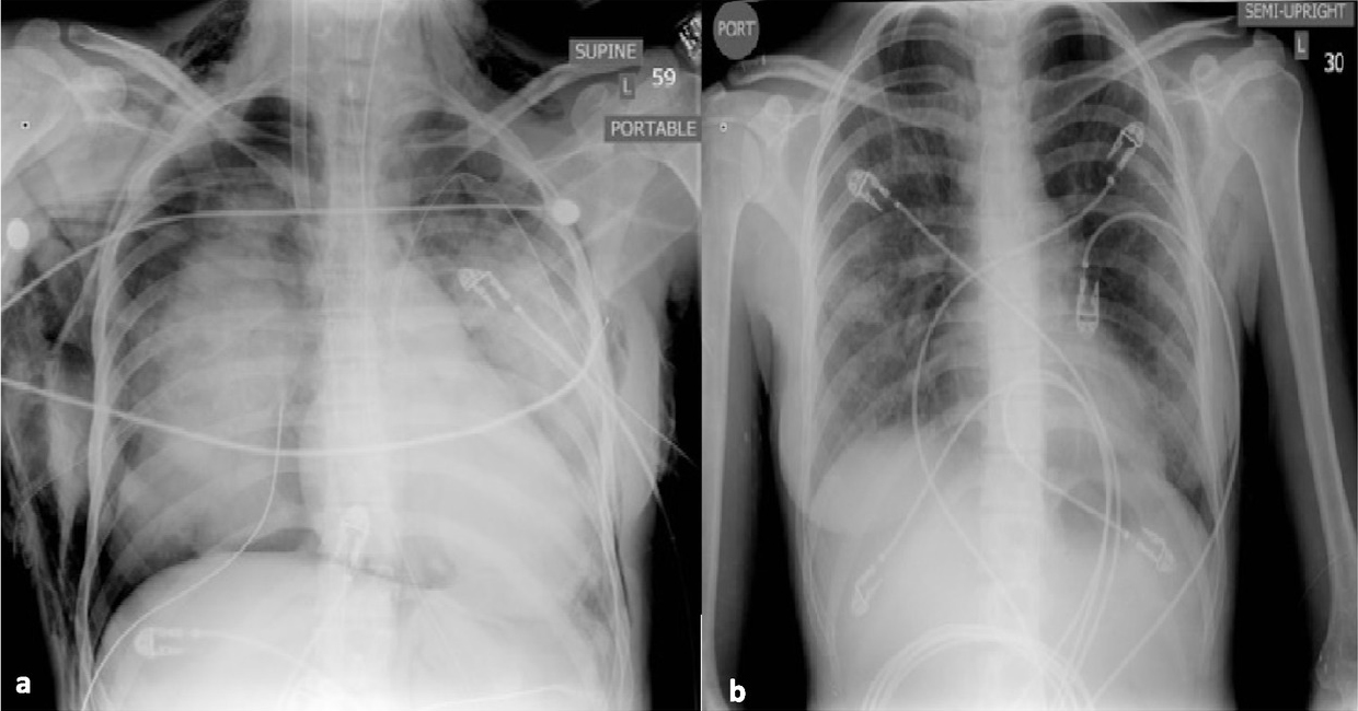
Fig. 2
a. Chest x-ray AP view following bilateral chest tube placement that shows bilateral patchy opacification with moderate right pneumothorax and diffuse subcutaneous emphysema along the thorax and neck. b. Chest x-ray AP view, day 9 of hospitalization after removal of bilateral chest tubes.