
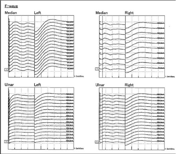
Fig. 1
Electroneurography revealed proximal conduction blocks detected by the absence of F waves on both median and ulnar nerves bilaterally.
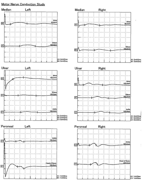
Fig. 2
Electroneurography was performed and revealed axonal loss in all motor nerves (the amplitude of compound muscle action potential [CMAP] was reduced more than 70%).
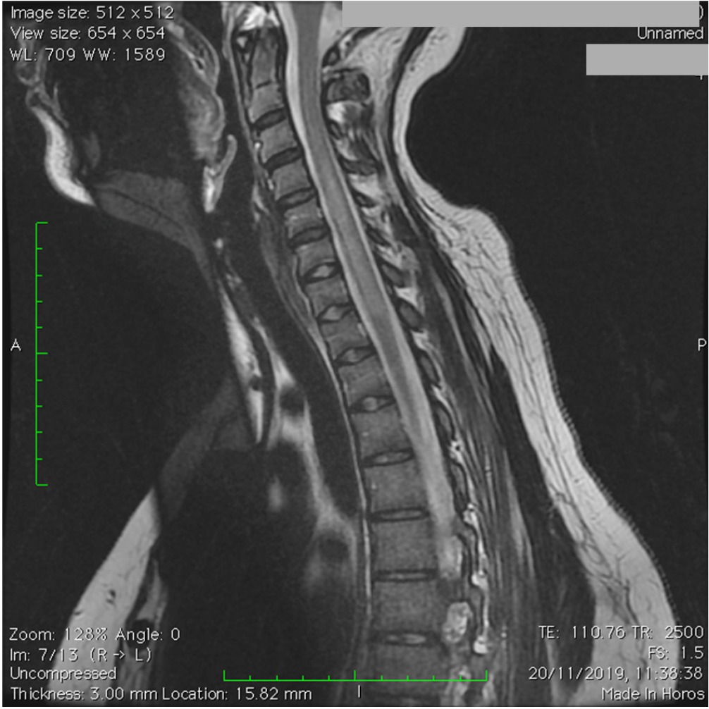
Fig. 3
Sagittal T2 weighted cervical MRI revealing a hyper-intense longitudinally extensive spinal cord lesion
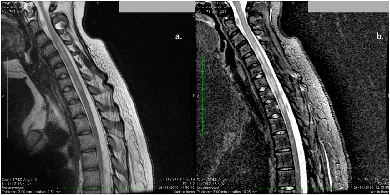
Fig. 4
Sagittal a. T2 weighted; b. Short tau inversion recovery (STIR) cervical MRI revealing a hyperintense longitudinally extensive spinal cord lesion