
Table 1
Results of relevant blood tests (on admission day)
| Test | Result | Normal range |
|---|---|---|
| White cell count | 9.8 109/L | 4-11 109/L |
| Neutrophils | 8.8 109/L | 2-7.5 109/L |
| Lymphocytes | 0.2 109/L | 1-4 109/L |
| Platelets | 62 109/L | 135-450 109/L |
| C-reactive protein | 643mg/L | 0-5 mg/L |
| Creatinine | 232 μmol/L | 45-84μmol/L |
| Urea | 19.7mmol/L | 2.5-7.8 mmol/L |
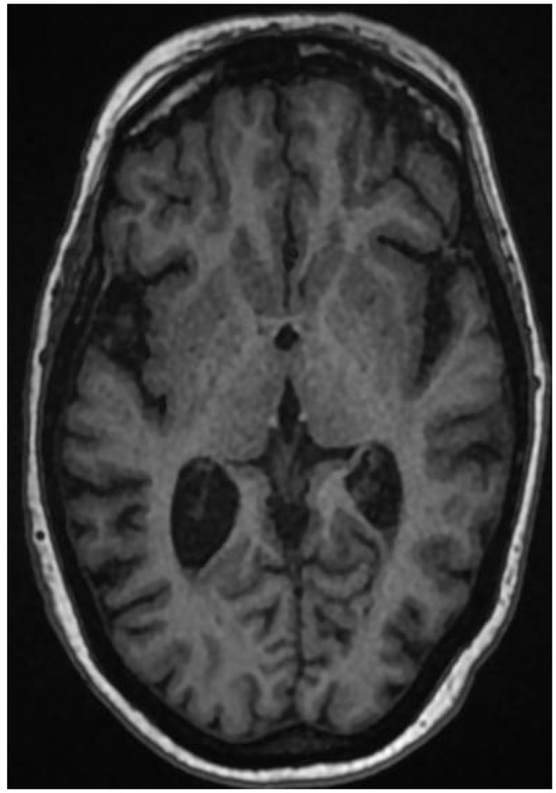
Fig. 1
Non contrast MRI brain on admission (5/10/2018) showing debris in the posterior horns of both the ventricles.
Table 2
LP results (admission day)
| CSF appearance | CSF results | |
|---|---|---|
| White cell count | 15 680 106/L | |
| Polymorphs | 95% | |
| Lymphocytes | 5% | |
| Turbid | Red blood cells | 220 106/L |
| Protein | 4.26g/L | |
| Glucose | 0.4mmol/L | |
| Serum glucose | 7.0mmol/L | |
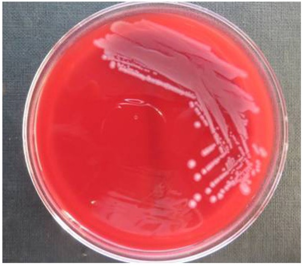
Fig. 2
E. coli colonies in blood agar.
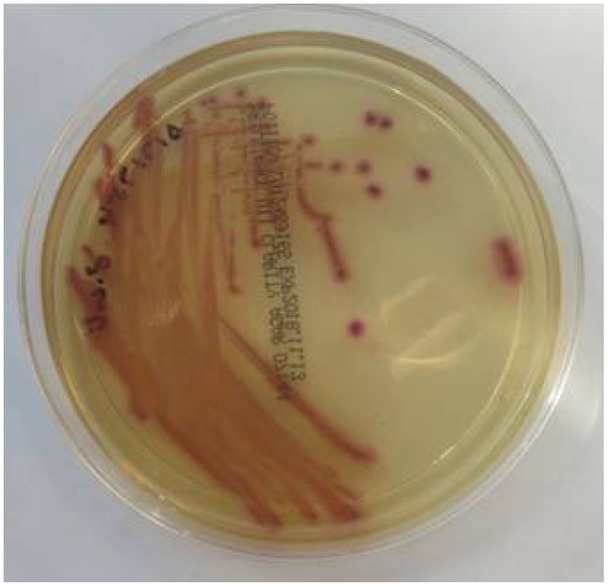
Fig. 3
E. coli colonies in chromogenic agar.
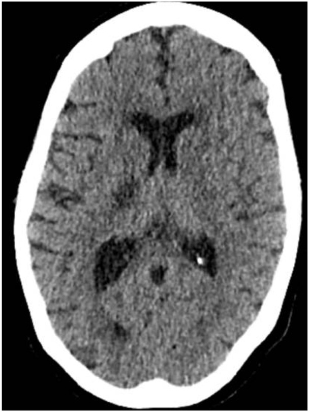
Fig. 4
Non contrast CT brain (day 7 post admission, 12/10/2018) showing a low attenuation area in the posterior limb of the right internal capsule, in keeping with a recent infarct.
Table 3
LP results (day 11)
| CSF appearance | CSF results | |
|---|---|---|
| White cell count | 14 106/L | |
| Polymorphs | 30% | |
| Lymphocytes | 70% | |
| Clear and colourless | Red blood cells | 1 106/L |
| Protein | 0.55g/L | |
| Glucose | 4.1mmol/L | |
| Serum glucose | 6.8mmol/L | |
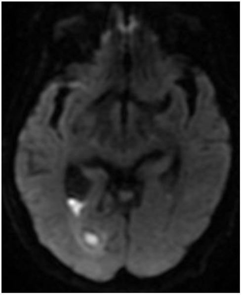
Fig. 5
MRI brain with contrast (Day 27, 2/11/2018) showing diffusion restriction in the posterior horn of the right lateral ventricle with trapped occipital horn.
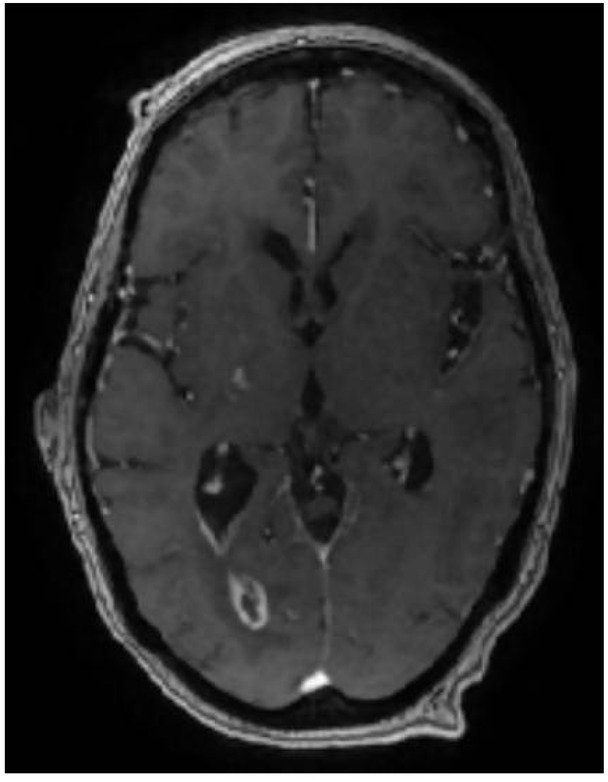
Fig. 6
MRI brain with contrast (Day 41, 16/11/2018) showing mild reduction in the size of the ring-enhancing lesion in the occipital horn of the right lateral ventricle.