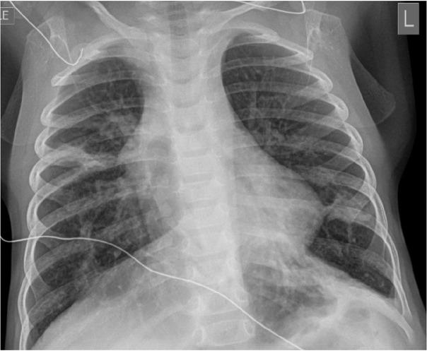
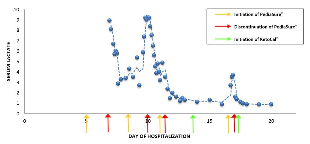
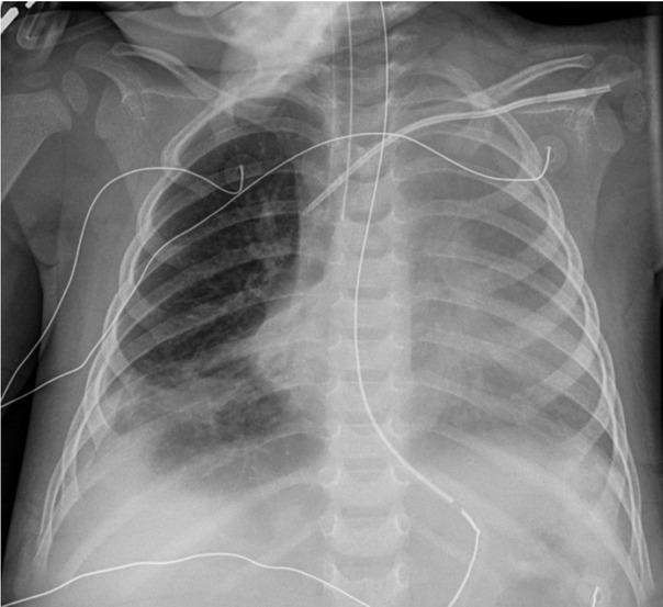
Have a personal or library account? Click to login
Pyruvate Dehydrogenase Complex Deficiency: An Unusual Cause of Recurrent Lactic Acidosis in a Paediatric Critical Care Unit Cover

Pyruvate Dehydrogenase Complex Deficiency: An Unusual Cause of Recurrent Lactic Acidosis in a Paediatric Critical Care Unit

By: Neha Gupta and  Chrystal Rutledge  
Open Access
|May 2019

Figures & Tables

Fig. 1

Chest radiograph on admission. The image shows pulmonary hyperexpansion with interstitial prominence and areas of patchy density which may be due to a viral airway disease with areas of atelectasis.

Fig. 2

Change in lactate associated with introduction of feeds. The figure describes an increase in lactate after each time the patient is started on PediaSure® feeds. The level decreased to normal after discontinuation of PediaSure®. The figure also describes no change in lactate level after starting the patient on KetoCal® feeds.

Fig. 3

Chest radiograph on hospital day 13. The image shows generalized opacification of the left hemithorax with no mediastinal shift. This could represent a combination of pleural fluid and atelectasis. Persistent right lower lobe atelectasis is seen along with right pleural fluid.

Pertinent laboratory results of the patient

LabsValues
Time of collectionpHpCO2 (mmHg)pO2 (mmHg)HCO3 (mmol/L)Serum lactate (mmol/L)
2 hours prior to respiratory arrest (Hospital Day 6) (Capillary gas)7.42228114.28.9
Blood1 hour after respiratory arrest (Hospital Day 6) (Capillary gas)7.1241.512513.58.1
gases36 hours after initiation of Pediasure® (Hospital Day 8) (Venous gas)7.3151.145.725.49.3
After development of ARDS (Hospital Day 13) (Arterial gas)7.42525533.51.3
Serum pyruvate level (mg/dL)2.42 (Reference range: 0.3-1.5)
Blood lactate to pyruvate ratio14.87
Activated PDC level (nmol/min/mg protein)0.35 (Reference range: 1.2-6.52)
Skeletal muscle biopsyPDC/E3 ratio0.4 (Reference range: 0.82-4.54)
Pyruvate dehydrogenase and mitochondrial complex panelPositive for pyruvate dehydrogenase alpha 1 (PDHA1) gene mutation
DOI: https://doi.org/10.2478/jccm-2019-0012 | Journal eISSN: 2393-1817 | Journal ISSN: 2393-1809
Language: English
Page range: 71 - 75
Submitted on: Mar 28, 2019
Accepted on: Apr 28, 2019
Published on: May 13, 2019
Published by: University of Medicine, Pharmacy, Science and Technology of Targu Mures
In partnership with: Paradigm Publishing Services
Publication frequency: 4 issues per year

© 2019 Neha Gupta, Chrystal Rutledge, published by University of Medicine, Pharmacy, Science and Technology of Targu Mures
This work is licensed under the Creative Commons Attribution-NonCommercial-NoDerivatives 4.0 License.