
Table 1
Specific primers used for detection of honey bee pathogens
| No. | Pathogen | Primer sequence (5′-3′) | Amplicon size (bp) | Annealing temp (°C) | Reference | |
|---|---|---|---|---|---|---|
| 1 | SBV | F: ACCAACCGATTCCTCAGTAG R: CCTTGGAACTCTGCTGTGTA | 487 | 57 | Grabensteiner et al., 2001 | |
| 2 | ABPV | F: TTATGTGTCCAGAGACTGTATCCA R: GCTCCTATTGCTCGGTTTTTCGGT | 901 | 55 | Benjeddou et al., 2001 | |
| 3 | CBPV | F: AGTTGTCATGGTTAACAGGATACGAG R: TCTAATCTTAGCACGAAAGCCGAG | 455 | 55 | Ribiere et al., 2002 | |
| 4 | Virus | DWV | F: TCATCTTCAACTCGGCTTTCTACG R: CGAATCATTTTCACGGGACG | 479 | 62 | Lee et al., 2005a |
| 5 | BQCV | F: TGGTCAGCTCCCACTACCTTAAAC R: GCAACAAGAAGAAACGTAAACCAC | 701 | 55 | Benjeddou et al., 2001 | |
| 6 | KBV | F: GATGAACGTCGACCTATTGA R: TGTGGGTGGCTATGAGTCA | 415 | 50 | Stoltz et al., 1995 | |
| 7 | IAPV | F: GATTTGAGAGATGTATTTCCTTCTGCGG R: ACACTTGCGTTGGTCCTGAATGTTAATGG | 725 | 52 | This study | |
| 8 | Bacteria | Paenibacillus larvae | F: GTGTTTCCTTCGGGAGACG R: CTCTAGGTCGGCTACGCATC | 232 | 55 | Lee et al., 2004 |
| 9 | Melissococcus plutonius | F: AAGAGTAACTGTTTTCCTCG R: AAACCTTATCTCTAAGGCGT | 583 | 52 | Ha et al., 2005 | |
| 10 | Ascosphaera apis | F: GGCTGTAGGGGGGAACCAGGA R: CGGGTGGTCGTTTCCAGCCTC | 995 | 62 | Lee et al., 2005b | |
| 11 | Fungus | Aspergillus flavus | F: ATCGGGCGGTGTTTCTATG R: ACCGGGCTATTTAAGGGCCG | 311 | 55 | Lee et al., 2004 |
| 12 | Nosema sp. | F: CTGCCTGACGTAGACGCTAT R: CTTCGATCCTCTAGCTTACG | 592 | 50 | Yoo et al., 2008 | |
| 13 | Parasite | Acarapis woodi | F: CAGTAGGGCTAGATATCGATACCCGAGCTT R: TGAGCTACAACATAATATCTGTCATGAAGA | 247 | 55 | This study |
| 14 | Apocephalus borealis | F: GTACACCTATACATTGGGTTCGTACATTAC R: GAGRGCCATAAAAGTAGCTACACC | 500 | 57 | This study | |
[i] Abbreviation: SBV- sacbrood virus; ABPV- acute bee paralysis virus; CBPV- chronic bee paralysis virus; DWV- deformed wing virus; BQCV- black queen cell virus; KBV- Kashmir bee virus; IAPV- Israeli acute bee paralysis virus.
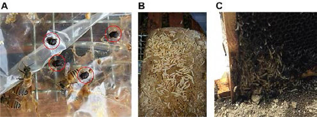
Fig. 1
Infestation of small hive beetle in honeybee hives. Aethina tumida in apiaries were diagnosed through the observation of the presence of adults (A), and larvae in supplied pollen (B) and honeycomb of infested hive (C).
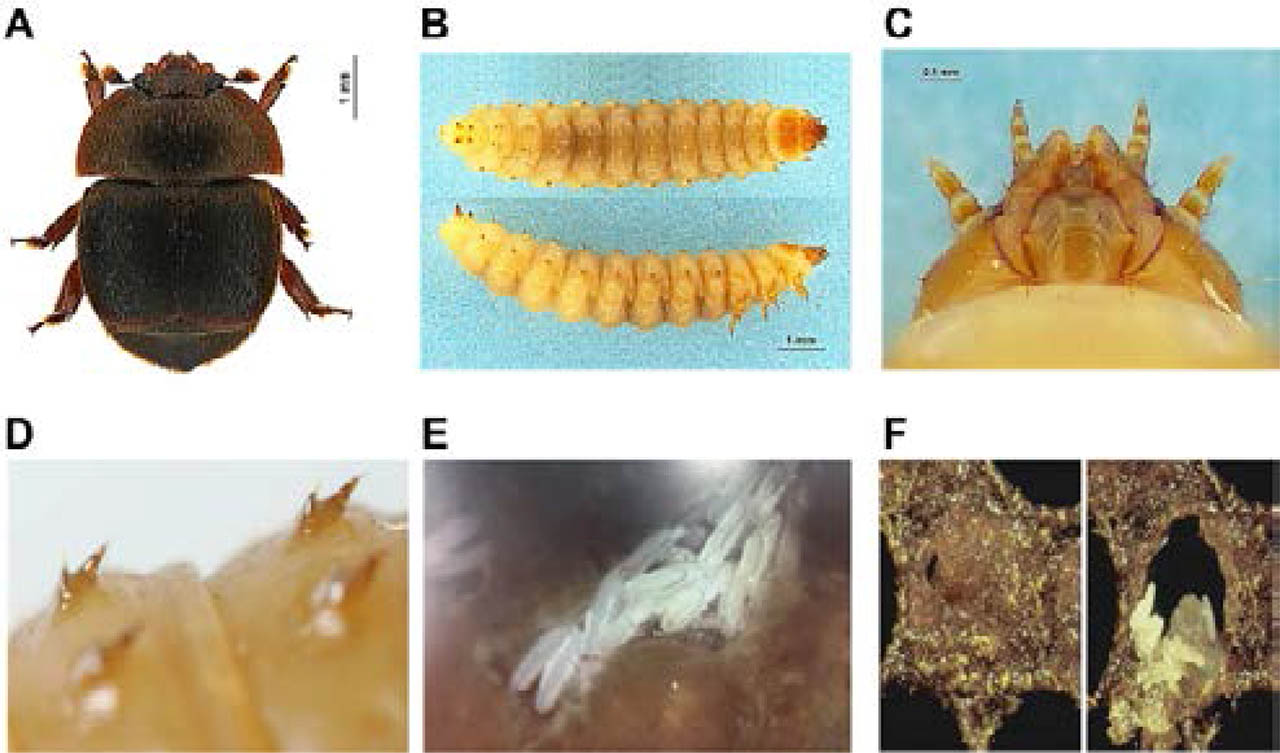
Fig. 2
Identification of small hive beetle based on morphological characteristics. Appearance of adults observed with 5~7 mm × 3~5 mm (length × width) of body size, dark brown colour, one pair of clubbed antennae on the head, and body shape of oval and dorsoventrally flattened (A). The larvae were characterized by three pairs of legs located close to the head, two rows of dorsal spines, two larger paired spines on the posterior segment of the dorsum, and body size was around 10 mm in length (B-D). Eggs of SHB were seen in capped bee brood with pearly-white colour and size of around 1.4 mm × 0.26 mm (length × width) (E and F).
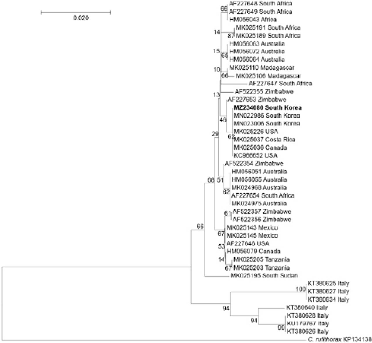
Fig. 3
Phylogenetic tree of mitochondrial cytochrome c oxidase I (COI) gene sequences from small hive beetle (SHB). Neighbor-joining phylogenetic tree was created based on mitochondrial COI gene of SHB with 1000 bootstrap iterations in MEGA7. Aethina tumida detected in this study with NCBI accession number MZ234080 was written in bold. Other reference strains of A. tumida with NCBI accession numbers and country names are shown. Cratonura rufithorax was used as an outgroup. Number above line represents bootstrap percentages.
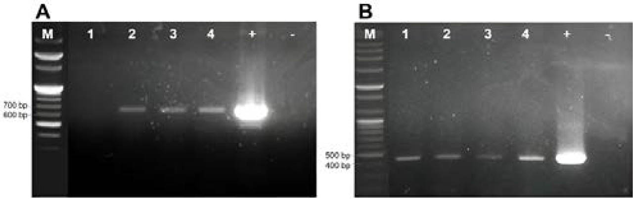
Fig. 4
Detection of honeybee pathogens in small hive beetle (SHB). Positive results of black queen cell virus (A) and deformed wing virus (B) detection were confirmed with expected band, 701 bp and 479 bp long, respectively, in electrophoresis. Lanes 1 to 4 were for four SHB samples. “M” is a 100-bp DNA marker. “+” and “-” are positive using recombinant DNA and negative control without DNA template, respectively.