
Table 1:
Comparative analysis of recent BT classification studies
| Study | Methodology | Dataset | Key contributions | Limitations |
|---|---|---|---|---|
| Shib et al [1] | VGG16-based CNN | Kaggle Brain MRI | Demonstrated pretrained CNNs improve classification accuracy | No temporal/sequential modeling; single-step classification |
| Hafeez et al. [2] | CNN with custom layers | Public MRI dataset | Reinforced importance of convolutional feature extraction | No recurrent or sequential modeling explored |
| Manjunath et al. [6] | Fine-tuned deep learning models | Brain MRI Dataset | Pretrained architectures provide strong baseline | Extensive hyperparameter tuning required; no hybrid model |
| Khan et al. [4] | Hybrid-Net (DenseNet + SVM) | Public MRI dataset | Combined deep features with traditional classifiers | No ablation study; lacks temporal modeling |
| Saeedi et al. [8] | CNN + SVM | Public Brain MRI | Two-step pipeline (feature extraction + classification) | No end-to-end joint optimization |
| Bouhafra & El Bahi [9] | Systematic review | Multiple MRI datasets | Comprehensive survey of deep learning methods | No sequential modeling in proposed frameworks |
| Alrashedy et al. [10] | BrainGAN (GAN + CNN) | Public brain MRI | Synthetic data augmentation to address dataset size | No recurrent/temporal modeling |
| Tabatabaei et al. [11] | CNN + transformer | Brain MRI Dataset | Attention-based feature modeling improves interpretability | High computational cost; lacks explicit temporal modeling |
| Şahin et al. [12] | ViT | Brain MRI Dataset | Multiobjective optimization for efficient classification | Global attention may miss local sequential dependencies |
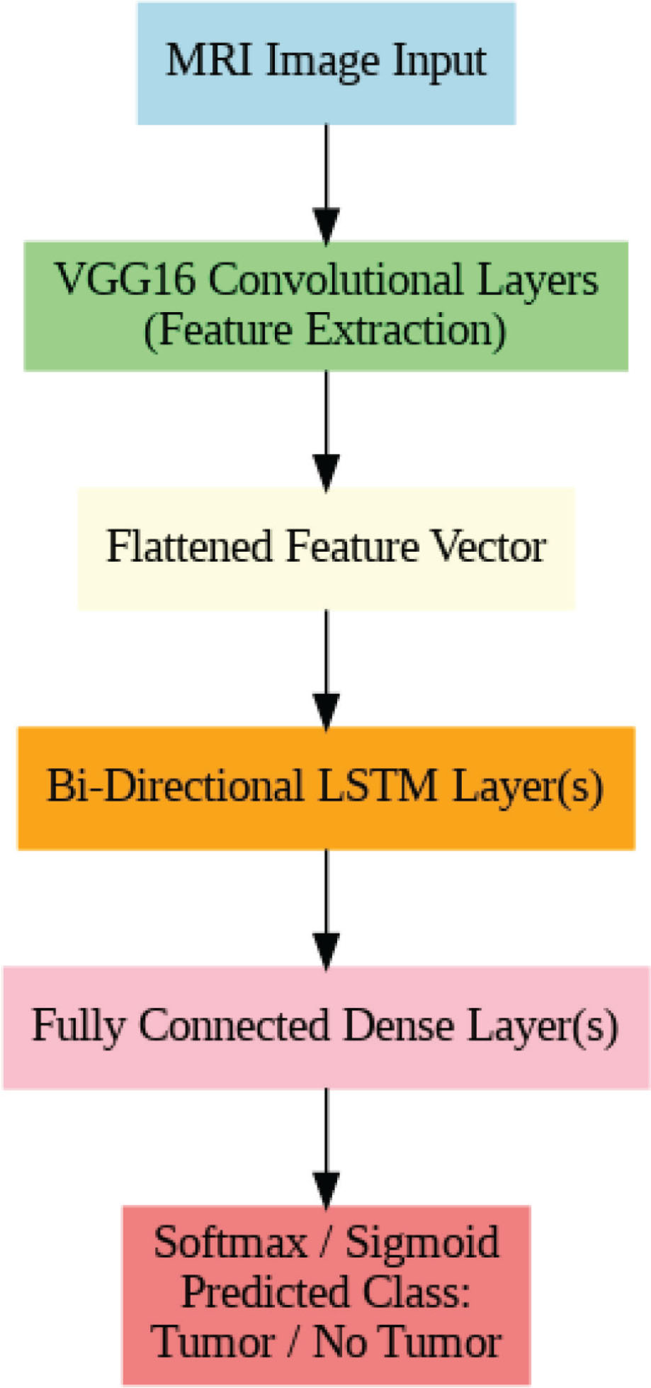
Figure 1:
Overview of the architecture. MRI, magnetic resonance imaging.
Table 2:
Hyperparameter tuning
| Hyperparameter | Final value |
|---|---|
| Learning rate | 0.0001 |
| Optimizer | Adam |
| Batch size | 32 |
| Epochs | 50 |
| BiLSTM units | 128 |
| Dropout rate | 0.5 |
Table 3:
Quantitative performance metrics
| Metric | Mean (%) | 95% CI (%) |
|---|---|---|
| Accuracy | 96.8 | ±0.5 |
| Precision | 97.1 | ±0.4 |
| Recall | 96.5 | ±0.6 |
| F1-score | 96.8 | ±0.5 |
| ROC-AUC | 0.985 | ±0.004 |
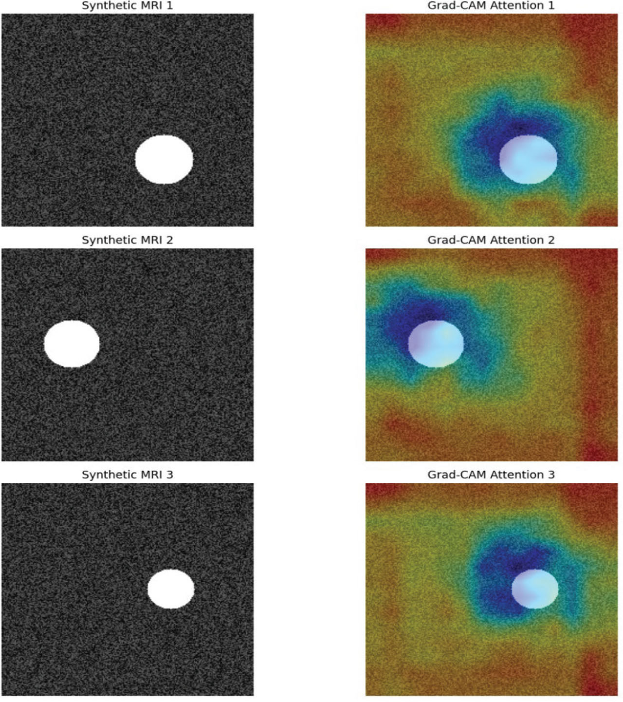
Figure 2:
Grad-CAM attention maps generated from the VGG16 encoder of the proposed VGG16-CAE + BiLSTM framework. BiLSTM, Bi-Directional Long Short-Term Memory; CAE, convolutional autoencoder.
Table 5:
Confusion matrix
| Predicted tumor | Predicted no tumor | |
|---|---|---|
| Actual tumor | 291 ± 5 | 9 ± 4 |
| Actual no tumor | 11 ± 3 | 289 ± 5 |
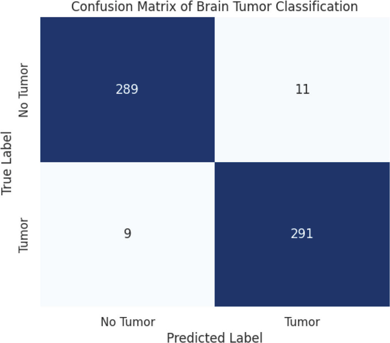
Figure 3:
Confusion matrix.
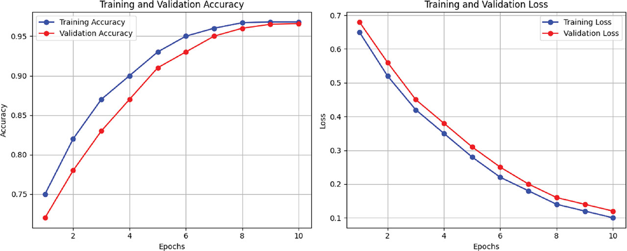
Figure 4:
Training and validation loss and accuracy curves for the VGG16-CAE + BiLSTM framework. BiLSTM, Bi-Directional Long Short-Term Memory; CAE, convolutional autoencoder.
Table 6:
Comparative evaluation of the proposed framework against baseline models
| Model | Accuracy (%) | Precision (%) | Recall (%) | F1-Score (%) | ROC-AUC |
|---|---|---|---|---|---|
| Standard VGG16 (pretrained CNN) | 91.2 ± 0.8 | 91.5 ± 0.7 | 90.8 ± 0.9 | 91.1 ± 0.8 | 0.943 ± 0.007 |
| ResNet50 (pretrained CNN) | 92.5 ± 0.7 | 92.7 ± 0.6 | 92.0 ± 0.8 | 92.3 ± 0.7 | 0.951 ± 0.006 |
| DenseNet121 (pretrained CNN) | 93.1 ± 0.7 | 93.5 ± 0.6 | 92.7 ± 0.7 | 93.1 ± 0.7 | 0.957 ± 0.005 |
| VGG16-CAE (without BiLSTM) | 94.3 ± 0.6 | 94.5 ± 0.5 | 94.0 ± 0.6 | 94.2 ± 0.5 | 0.970 ± 0.005 |
| Proposed VGG16-CAE + BiLSTM | 96.8 ± 0.5 | 97.1 ± 0.4 | 96.5 ± 0.6 | 96.8 ± 0.5 | 0.985 ± 0.004 |
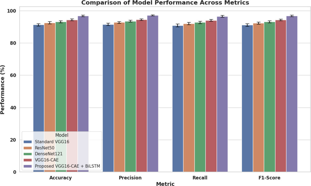
Figure 5:
Comparison of model performance across various models.
Table 7:
ROC analysis
| Model | ROC-AUC |
|---|---|
| VGG16 | 0.943 |
| ResNet50 | 0.951 |
| DenseNet121 | 0.957 |
| VGG16-CAE (without BiLSTM) | 0.970 |
| Proposed VGG16-CAE + BiLSTM | 0.985 |
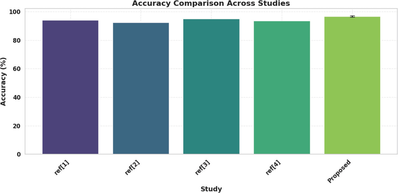
Figure 6:
Accuracy analysis.
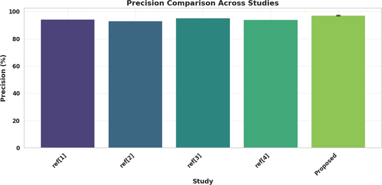
Figure 7:
Precision analysis.
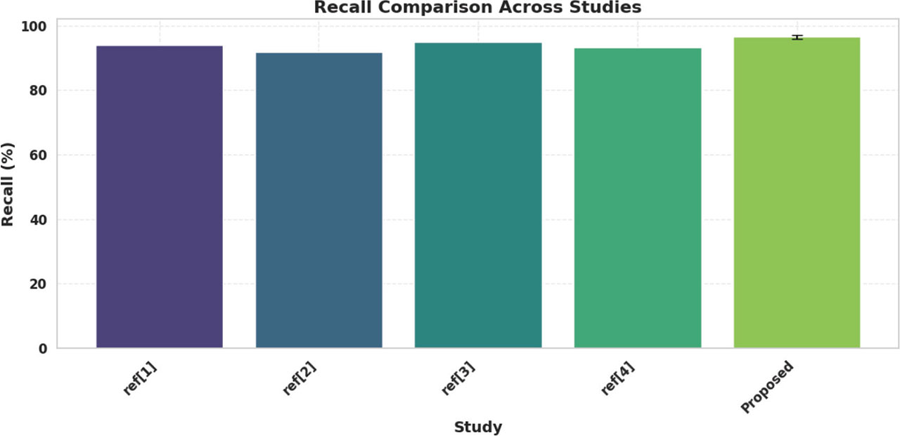
Figure 8:
Recall analysis.
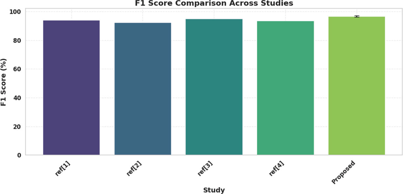
Figure 9:
F1 score comparison.
Table 8:
Comparative analysis of the proposed framework with recent literature
| Study | Methodology | Dataset | Accuracy (%) | Precision (%) | Recall (%) | F1-Score (%) |
|---|---|---|---|---|---|---|
| Shib et al. [1] | VGG16-based CNN | Brain MRI Kaggle Dataset | 94.0 | 94.3 | 93.8 | 94.1 |
| Hafeez et al. [2] | CNN with custom layers | Public MRI dataset | 92.5 | 93.0 | 91.8 | 92.4 |
| Manjunath et al. (2025) [6] | Fine-tuned deep learning models | Brain MRI Dataset | 95.1 | 95.3 | 94.8 | 95.0 |
| Saeedi et al. (2023) [8] | CNN + SVM Classifier | Public brain MRI Dataset | 93.7 | 94.0 | 93.2 | 93.6 |
| Proposed VGG16-CAE + BiLSTM | VGG16-based CAE + BiLSTM | Kaggle Brain MRI Dataset | 96.8 ± 0.5 | 97.1 ± 0.4 | 96.5 ± 0.6 | 96.8 ± 0.5 |