
Introduction
Gastrointestinal parasitic infections represent a major issue for equine health, affecting their welfare, productivity, and performance. These parasitoses are mainly caused by nematodes, cestodes, and protozoa, each capable of inducing a wide range of pathologies, from mild digestive disorders to severe conditions such as colic, gastric ulcerations, and even death (Alegre & Milano, 2020; Sazmand et al., 2020; Efremova et al., 2022; Attia & Omar, 2025).
Strongyles (including large strongyles such as Strongylus vulgaris and small strongyles, particularly cyathostomins) are the most concerning gastrointestinal parasites in equids due to their high prevalence and significant pathogenic potential. They are often responsible for the majority of digestive disorders and deterioration in body condition (Sotiraki et al., 1997; Tavassoli et al., 2010; Gehlen et al., 2020). Cestodes and certain protozoa may also cause intestinal lesions or severe diarrhea, particularly in young animals (Alegre & Milano, 2020; Sazmand et al., 2020; Efremova et al., 2022). The prevalence of these parasites varies by region, management practices, and animal age, but high infestation rates are frequently reported, highlighting the importance of regular monitoring and tailored prevention strategies (Efremova et al., 2022; Golshang et al., 2024). Furthermore, increasing anthelmintic resistance and insufficient consideration of parasitoses in diagnostic protocols complicate management, making close surveillance essential to preserve equine health (Alegre & Milano, 2020; Sazmand et al., 2020; Golshang et al., 2024).
In Algeria, horses play a significant cultural and economic role. However, epidemiological data on equine parasitic infections remain scarce, particularly in the semi-arid region of Constantine. This lack of knowledge obscures the regional prevalence of parasites and suggests that even horses kept under rigorous breeding protocols, such as those at the Eastern Equestrian Club, can harbor large and diverse parasite populations. To address this gap, the present study aimed to characterize the parasitological status of these horses by combining qualitative (fecal flotation) and quantitative (fecal egg counting using the McMaster method) techniques.
Material and Methods
Sample collection and preparation
Twenty fresh fecal samples were collected from clinically healthy Arabian and Thoroughbred horses (Equus caballus) aged 3 – 15 years (juveniles 3 – 5 yr, n = 6; adults 6 – 10 yr, n = 9; seniors >10 yr, n = 5; 12 males and 8 females) at the Eastern Equestrian Club, Constantine, Algeria (36°21′N, 6°36′E). Sampling occurred between October 2023 and April 2024. This extended period was designed to cover contrasting autumn–winter seasons and capture seasonal variation in egg shedding; it also reflected logistical constraints related to animal availability and the application of strict sample-quality criteria that led to the exclusion of several specimens.
Samples were retrieved immediately following natural defecation using disposable gloves to prevent cross-contamination. Approximately 50 – 100 g of feces was collected from the interior of each bolus to minimize environmental contamination, transferred into sterile, labeled polyethylene bags, and refrigerated at 4°C. All samples were transported to the Laboratory of Parasitology at the Institute of Veterinary Sciences, University of Constantine 1, within 4 h of collection and processed within 24 h to preserve egg morphology and viability. Prior to laboratory processing, each sample underwent macroscopic examination under standard lighting to identify grossly visible parasites, including adult stages, cestodes, proglottids, or larvae. Samples exhibiting excessive desiccation, substantial bedding contamination, or abnormal physical consistency were excluded from subsequent analyses. All horses were enrolled in a routine prophylactic anthelmintic program (ivermectin or benzimidazoles); the last treatment had been administered at least 8 – 12 weeks prior to sampling.
Parasitological analysis
The study combined two complementary analytical approaches for a comprehensive assessment of parasitic status. The flotation method, a widely used qualitative coproscopic technique (Thienpont et al., 2003), is based on the principle of concentrating parasitic elements by density separation. Feces are mixed with a dense solution (density > 1.2), allowing, under centrifugation, the sedimentation of debris at the bottom of the tube while parasitic elements migrate to the surface. The standardized protocol consists of homogenizing 5 g of feces in tap water, followed by filtration through a sieve (250 μm). After centrifugation of the filtrate (2000 ×g, 5 min), the supernatant is discarded, and the pellet is resuspended in a saturated NaCl solution (40 g/100 mL, density = 1.2). A second centrifugation is performed under the same conditions, followed by adding solution until a convex meniscus forms, and the sample is then covered with a coverslip to trap parasitic elements. After 5 minutes of rest, the coverslip is removed and examined under a microscope (100 – 400×) to identify parasites. This method, reproducible and sensitive, is particularly suitable for diagnosing light infestations.
For precise quantification of parasite excretion, the modified McMaster technique was used as described by Taylor et al. (2016). It involves diluting 3 g of feces in 42 mL of distilled water, then homogenizing and filtering through double gauze (0.15 mm). The filtrate is centrifuged (1500 ×g, 5 min), and the pellet is resuspended in a saturated solution (density = 1.2) to obtain the initial volume. An aliquot is loaded into the chambers of a McMaster counting slide (0.15 mL/chamber), and oocysts are counted after 5 minutes of sedimentation under a microscope (100× magnification). The number of oocysts per gram of feces (EPG) is calculated using the formula:
where N represents the total number of oocysts counted in both chambers, 45 mL is the total suspension volume, 0.3 mL is the combined chamber volume, and 3 g is the weight of feces analyzed.Morphological identification
Identification of parasitic eggs was based on the analysis of their distinctive morphological characteristics, including size, shape, color, and the possible presence of specific structures such as a micropyle. Examination was performed using a light microscope equipped with a calibrated micrometer, consisting of two essential components (Thienpont et al., 2003): an objective micrometer (a slide graduated in 10 μm divisions) and an ocular micrometer (an eyepiece with a graduated scale that varies by model).
Precise measurement of parasitic structures was performed using the formula:
Data analysis and interpretation
Quantitative results were interpreted according to the classification system established by Euzéby (1981) and Ambrosi (1995), which defines five levels of infestation based on EPG values. This rigorous methodological approach, combining validated qualitative and quantitative techniques, enabled a comprehensive and reliable evaluation of the parasitic status of the studied horses.
For statistical analysis, data were processed using R software (v4.2.1). After checking normality (Shapiro-Wilk test), comparisons were performed using Student's t-test (normal data) or Mann-Whitney test (non-parametric data). For multiple comparisons, a one-way ANOVA followed by Tukey's post-hoc test was applied. Correlations were assessed using Pearson's test (significance threshold p < 0.05). Parasitic prevalence was calculated as [Prevalence = (positive samples/total) × 100], with 95 % confidence intervals. Differences in prevalence were tested using Chi-square or Fisher's exact test (for small sample sizes).
Ethical Approval and/or Informed Consent
All experimental procedures involving animals were reviewed and approved by the Institutional Animal Ethics Committee of the University of Constantine 1. Animal husbandry and experimental conditions complied with relevant national regulations and institutional policies for the care and use of animals, in accordance with current equid welfare guidelines (Waran, 2007; Department for Environment, Food and Rural Affairs, 2017; Olczak & Tomczyk-Wrona, 2022; Waran & Evans, 2024).
Results
Analysis of the prevalence of parasitic entities
Macroscopic examination of fecal samples did not detect visible parasitic elements (larvae or adults). However, microscopic analysis revealed an overall prevalence of parasitic infestation of 50 %, with 10 of 20 samples containing parasitic eggs or oocysts. This moderate prevalence suggests good effectiveness of the sanitary program implemented in the studied stable, while also indicating the persistence of certain infestations despite prophylactic measures.
The distribution of identified parasites (Fig. 1) shows a clear predominance of strongyles, detected in 60 % of positive cases (6/10). Other observed parasites included Strongyloides westeri (40 %, 4/10) and Habronema spp. (30 %, 3/10), Eimeria leuckarti (20 %, 2/10), and Parascaris spp. (10 %, 1/10). This parasitic diversity, although dominated by nematodes, highlights the need for a broad diagnostic approach including protozoa.
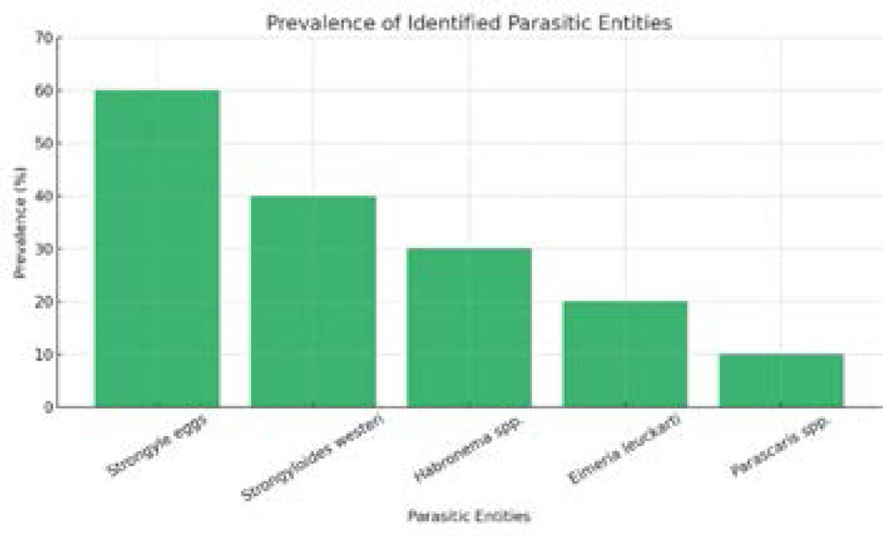
Fig. 1.
Prevalence of parasitic entities identified in fecal samples.
Morphological analysis of the parasite
Detailed morphological analysis of parasitic elements, illustrated in Figure 2, showing typical strongyle eggs (112.7 μm), enabled precise identification of different taxa. Systematic micrometric measurements, conducted according to the protocol of Thienpont et al. (2003), were a key factor in differentiating parasitic species.
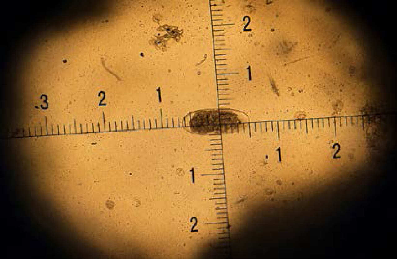
Fig. 2.
Strongyle eggs. Magnification ×10; 11.5 divisions × 9.8 μm (magnification index) = 112.7 μm.
Quantitative assessment
Quantitative analysis of eggs per gram of feces (EPG) across the 10 samples revealed a heterogeneous distribution of parasitic load (Fig. 3). Values ranged from 30 to 380 EPG, with infestation intensity predominantly low according to Ambrosi's classification (1995): 80 % of samples (n=8) were below 200 EPG, with individual values ranging from 30 to 104 EPG. Two subjects (20 %) exhibited light loads (200 – 380 EPG; samples 9 and 10), suggesting notable individual variation.
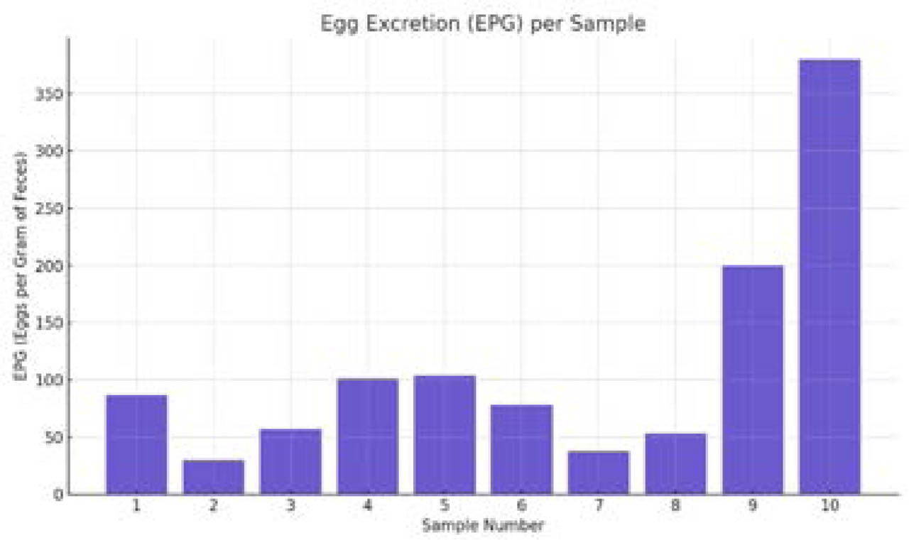
Fig. 3.
Distribution of parasitic excretion values (eggs per gram of feces - EPG) in analyzed samples.
This distribution with a concentration of low values among 80 % of samples (30 to 104 EPG) and two markedly higher cases (200 and 380 EPG), may reflect differences in parasite exposure among individuals, variability in immune response, or differential effectiveness of control measures. Although infestation remained generally low (median = 82.5 EPG), the presence of dispersed values (30 – 104 EPG; 200 – 380 EPG) highlights the importance of an individual-based approach in assessing parasitic status. The tow elevated values (200 and 380 EPG) although limited to minority of samples, warrant further investigation to rule out specific risk factors.
Discussion
Strongyle eggs were the most frequently detected parasites in this study, accounting for 60 % of positive fecal samples. This finding is consistent with numerous epidemiological data identifying strongyles as the primary gastrointestinal nematodes in equines, even in well-managed populations (Hinney et al., 2011; Rehbein et al., 2013; Jürgenschellert et al., 2022; Molento et al., 2024). Prevalence rates consistently exceeding 50 % in various geographical settings (Rehbein et al., 2013; Negash et al., 2021; Mulaw Berihun et al., 2024) highlight the remarkable environmental resilience of their free-living life stages and their ability to persist despite routine anthelmintic treatments.
Age and sex are recognized host factors modulating parasite susceptibility. In this cohort, juveniles (3 – 5 yr) tended to show higher shedding of age-associated parasites (S. westeri, Parascaris spp.), consistent with the literature (Relf et al., 2013; Boelow et al., 2023). No significant sex-related difference was detected, likely owing to the limited sample size.
Moreover, Strongyloides westeri was detected in 40 % of cases, an unusually high frequency compared with the typically reported range of 0.5 % to 8 % (Lyons & Tolliver, 2004; Relf et al., 2013; Elghryani et al., 2023). This particularity may be explained by the presence of young animals or environmental conditions favorable to its development. Similarly, Habronema spp., observed in 30 % of cases, exhibit a variable geographical distribution, with higher prevalence in environments conducive to vector-borne transmission, such as those involving flies.
The presence of Eimeria leuckarti in 20 % of horses, although based on a limited sample size, is a notable finding that warrants special attention. This protozoan is the only species of the genus Eimeria recognized as pathogenic in equids, with a more pronounced clinical impact in young animals, particularly foals. These may develop severe diarrhea and enteritis in some cases, although most infections remain asymptomatic or of limited clinical significance (Dubey & Bauer, 2018; Güleğen et al., 2016; Rehman et al., 2021). Its prevalence varies depending on region and management conditions, with higher rates observed in foals and in humid or poorly hygienic environments (Güleğen et al., 2016; Rehman et al., 2021). Although most studies report low oocyst excretion and absence of clinical signs in adults, pathogenic cases do occur, highlighting the need for increased vigilance, particularly in breeding systems housing young horses (Dubey & Bauer, 2018; Gorji et al., 2023).
These observations justify adopting an expanded diagnostic approach that includes not only helminths but also protozoa such as E. leuckarti to better prevent and manage digestive disorders in equids (De Souza et al., 2009; Dubey & Bauer, 2018; Rehman et al., 2021). Optimization of management practices, particularly regarding hygiene, feeding, and watering, could help reduce the prevalence of this parasite (Rehman et al., 2021).
Parascaris spp., detected in 10 % of cases, completes this parasitological profile. This parasite isß more common in foals, with prevalence ranging between 4 % and 22 % depending on the study (Lyons & Tolliver, 2004; Boelow et al., 2023; Elghryani et al., 2023; Molento et al., 2024). The diversity of identified parasitic species, combined with the frequency of co-infections, complicates health management, as each species presents distinct biological cycles and pathogenic impacts (Rehbein et al., 2013; Jürgenschellert et al., 2022). Therefore, an integrated strategy combining targeted deworming, improved hygiene, and appropriate management of feeding and watering is essential for effective control (Elghryani et al., 2023; Molento et al., 2024).
The intensity of infestation was evaluated according to the scale proposed by Ambrosi (1995) and Euzéby (1981), which classifies infestations into different categories: less than 200 eggs per gram (EPG) corresponds to low infestation, 200 – 500 EPG to light infestation, 500 – 600 EPG to moderate infestation, 600 – 1000 EPG to high infestation, and above 1000 EPG to severe infestation. This quantitative approach, increasingly used in parasitology, allows better assessment of infestation severity and more precise adaptation of control measures (Kaplan & Nielsen, 2010; Nielsen, 2012). According to this classification, most infested horses (8 out of 10) exhibited low infestation, while two individuals (samples 9 and 10) showed light infestation (200 and 380 EPG, respectively). This distribution, characterized by a predominance of low-intensity cases, is consistent with the well-documented aggregated pattern of strongyle egg shedding in equine populations, where the majority of horses act as low shedders (Relf et al., 2013; Nielsen et al., 2014). These results suggest relatively effective parasite control in the studied stable. Low to light excretion levels reflect well-managed antiparasitic practices, including regular diagnostic testing such as coproscopy and adaptation of treatments based on results. These practices are recommended to limit excessive anthelmintic use and prevent the development of resistance (Stratford et al., 2014; Rendle et al., 2024; Beasley et al., 2025).
However, the persistence of multiple parasitic species indicates that the risk of reinfestation is not fully controlled. This situation may be explained by the resilience of parasite life cycles in the environment, in which eggs and larvae can persist for long periods in pastures, making complete eradication difficult even with rigorous management (Shrubb et al., 2025; Raza et al., 2019). It may also be attributed to the emergence of anthelmintic resistance, particularly in strongyles and Parascaris spp. Intensive and repeated use of the same drug classes has indeed promoted resistance development (Martin et al., 2024; Nielsen et al., 2014). Recent studies confirm resistance to several molecules, including fenbendazole, pyrantel, and even macrocyclic lactones, compromising treatment efficacy and complicating infestation management (Martin et al., 2024; Raza et al., 2019).
Conclusion
This study reveals a contrasted parasitological situation in horses at the Eastern Equestrian Club in Constantine. Although the over-all prevalence of infestations (50 %) and the intensity of parasitic excretion (mostly low according to Ambrosi and Euzéby classification) indicate generally effective sanitary control, the diversity of identified species highlights the persistence of non-negligible parasitic risks. The predominance of strongyles (60 % of positive cases), confirming their status as major parasites in equids, is accompanied by the notable presence of other parasitic species.
This diversity, combined with the occasional detection of light burdens (up to 380 EPG), highlights the limitations of current deworming protocols in the face of resilient parasite life cycles and possible anthelmintic resistance. The presence of E. leuckarti, although often asymptomatic in adults, calls for increased vigilance in more vulnerable foals.
These findings call for improved parasite management by combining regular diagnosis (including protozoa and helminths), targeted deworming, and the optimization of management conditions (hygiene, grazing). In the Algerian context, where equine parasitological data remain scarce, this study provides essential epidemiological insights to inform the adaptation of prevention strategies and emphasizes the need to monitor the emergence of resistance and strengthen biosecurity measures. Further investigations, including larger sample sizes and molecular analyses, would help refine these conclusions and optimize equine health in the region.