
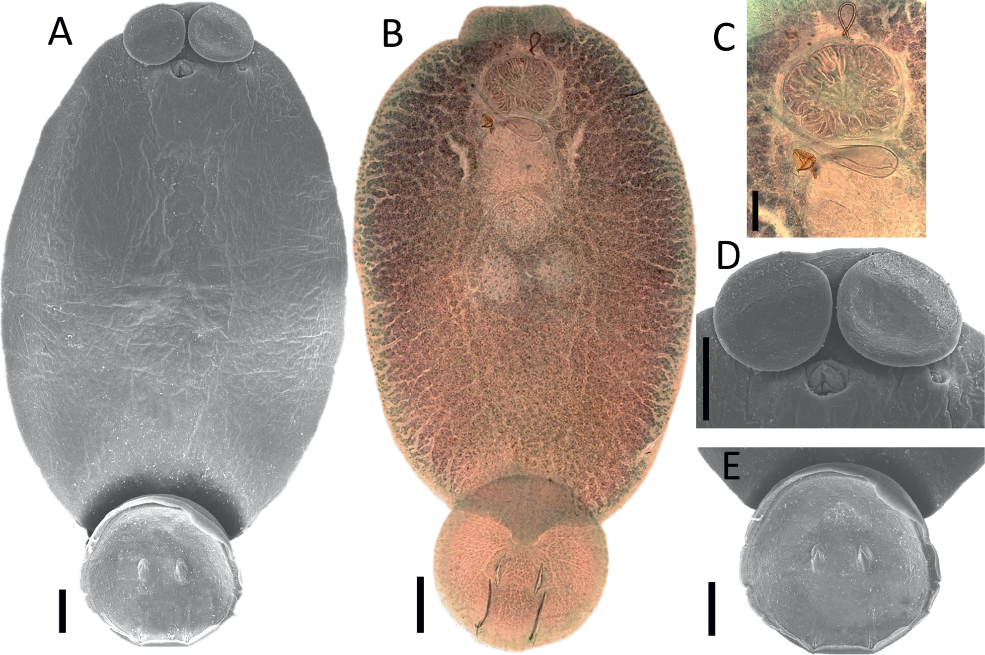
Fig. 1.
Neobedenia girellae, A) Photomicrograph of scanning electron microscopy (SEM) of an adult, ventral view; B) Photomicrograph of an adult specimen stained with Mayer’s paracarmine, dorsal view; C) Detail of the anterior end of a stained specimen showing a filamented egg; D) SEM of anterior end showing the attachment organs; E) SEM of posterior end showing the structure of the haptor. Scale bar = 200 μm
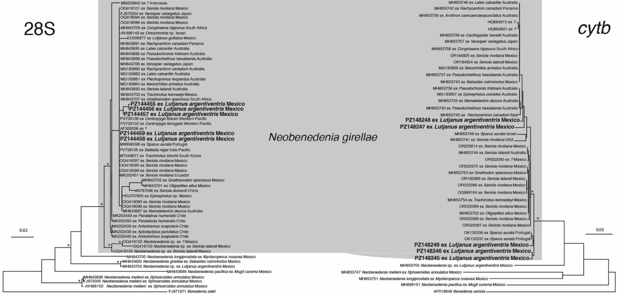
Fig. 2.
Maximum-likelihood phylogenetic trees (28S and cytb) showing the phylogenetic position of Neobenedenia girellae isolates from Lutjanus argentiventris in the Estero Urías, Mazatlán. Asterisks above nodes represent bootstrap support values higher than 80.