
Table 1.
Target gene and primer sets used for the detection of Echinococcus spp.
| Echinococcus spp. | Target gene | Primer | 5’-3’ | PCR Product | Reference |
|---|---|---|---|---|---|
| Echinococcus granulosus | nd1 | ND1-F | TGTTGCAGAGGTTTGCTGAT | 674 bp | UGene lab |
| ND1-R | ACGAACACGTGGTAATGTCG | ||||
| Echinococcus multilocularis | nd1 | EMND1-1F | TTGTTCTTTGTGTTACTGTAGG | 457 bp | Shang et al. (2019) |
| EMND1-1R | CTATACAGACATTGATTACCATAA | ||||
| Echinococcus equinus | nd2 | EEQND2-2F | CGTTAATTCACTGATACATTGTATCC | 551 bp | UGene lab |
| EEQND2-1R | CTCACACCAAGCACCTACAC |
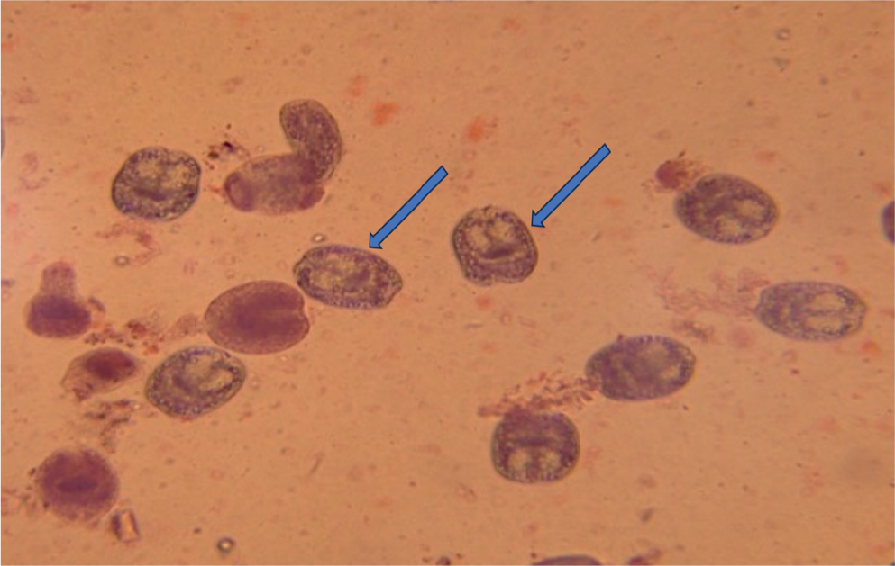
Fig. 1.
Viability of protoscolices using eosin stain (0.1 %), where stained is dead and unstained is live (blue arrow).
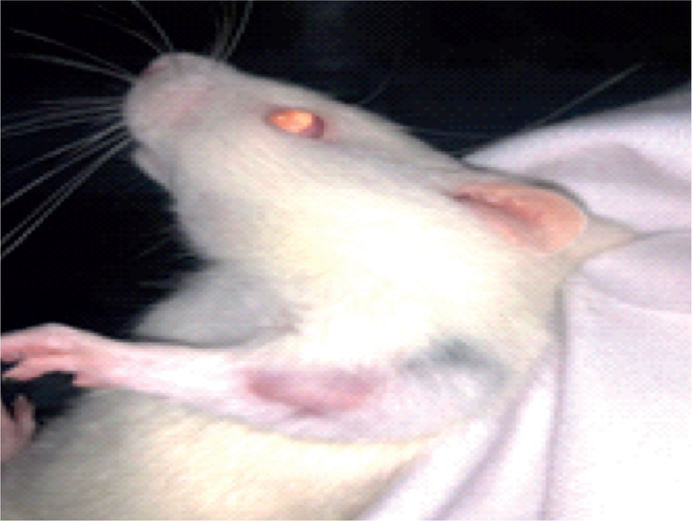
Fig. 2.
Casoni test with a hypersensitivity reaction (erythematous spot) on the upper left hand.
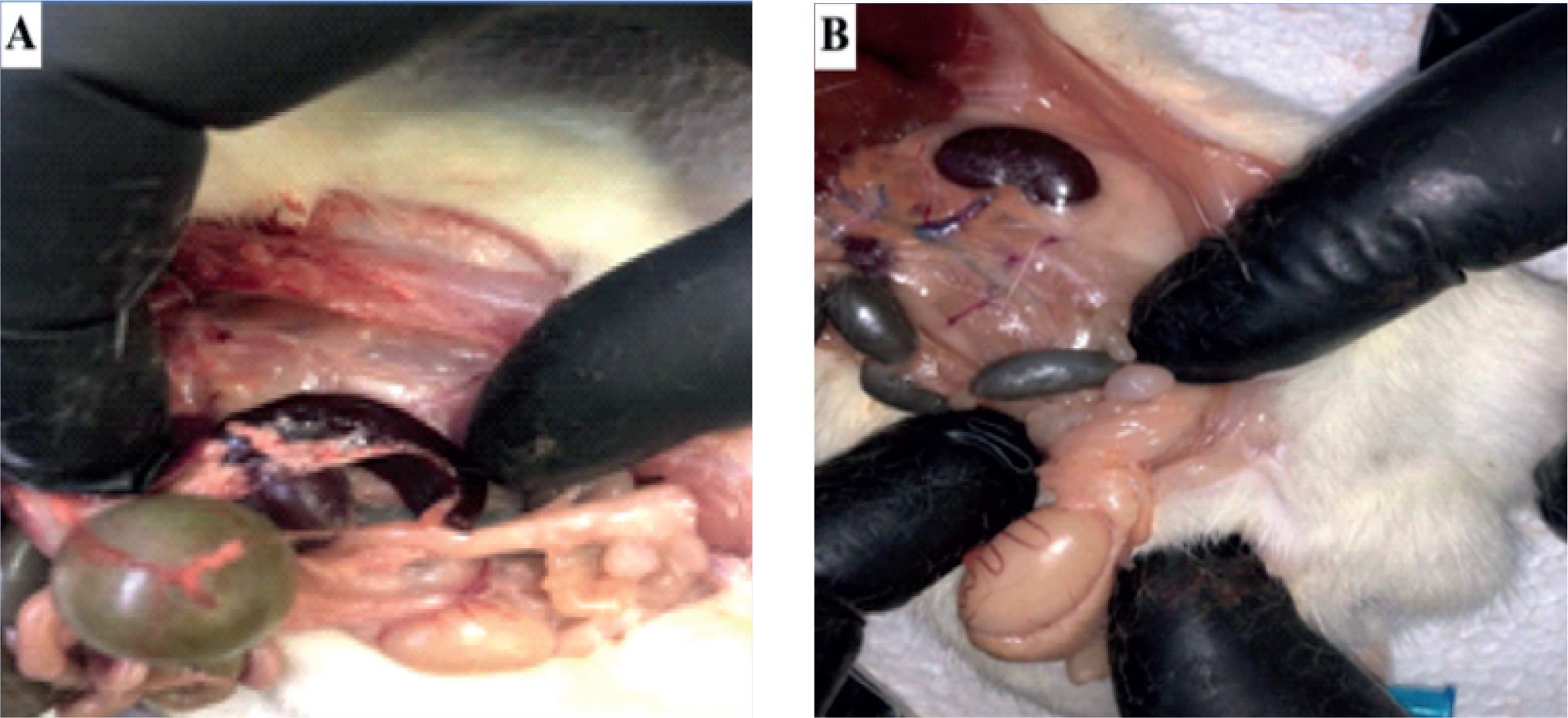
Fig. 3.
Cyst of hyadits in the viscera of the autopsied rats (A & B images).
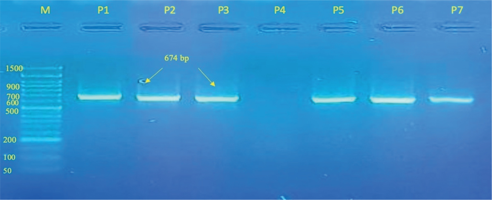
Fig. 4.
Gel electrophoresis showing the amplicon of the nd1 gene of Echinococcus granulosus from protoscolices samples. The DNA marker (M) is a 50-1500 bp ladder, and the six samples show a distinct 674 bp band.
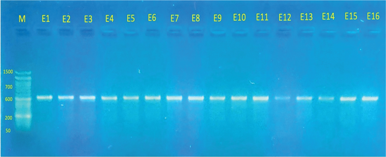
Fig. 5.
Gel electrophoresis showing the amplicon band of the nd1 gene of E. granulosus from the plasma samples; The DNA marker is a 50 to 1500 bp ladder, and the band size of sixteen samples is displayed as a distinct band at 674 bp.
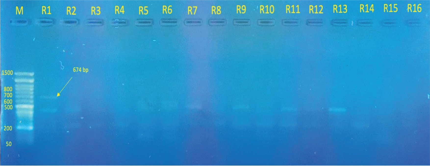
Fig. 6.
Gel electrophoresis showing the amplicon of the nd1 of E. granulosus from serum samples. The DNA marker (M) is a 50-1500 bp ladder, and only one sample (R1) displayed a distinct band at 674 bp. Other uncleared bands with lengths less than 500 bp may belong to DNA random fragments of Echinococcus in the bloodstream that are not specifically targeted.
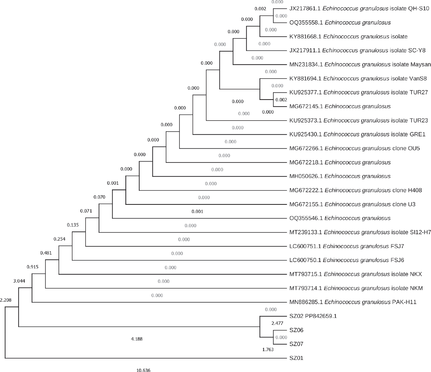
Fig. 7.
Evolutionary relationships of partial ND1 gene sequences from four isolates (sequenced; only one (SZ02) is recorded in the Gene Bank) belong to E. granulosus.