
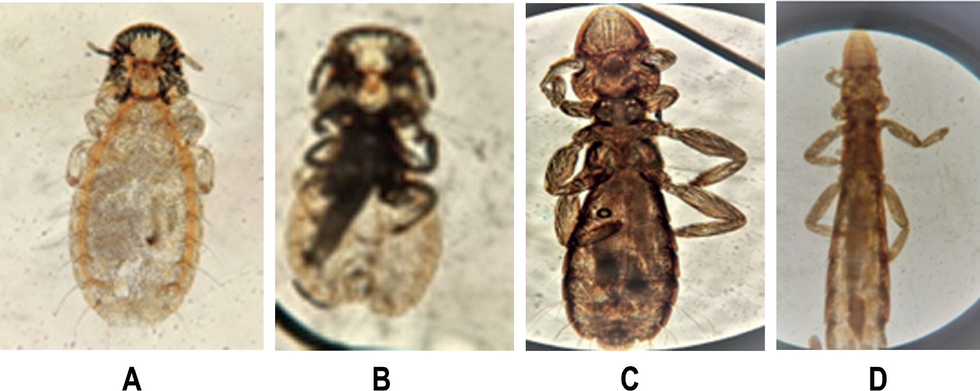
Fig 1.
The detected lice. (A) Ventral side of C. compar (10X); (B) Dorsal side of C. compar (5X); (C) Male of C. tschulyschman (10X); (D) Female of C. tschulyschman (10X).
Table 1.
Prevalence of ectoparasitic infestation in relation to sex of the pigeons.
| Gender | Infected, N (%) | Uninfected, N (%) | P-value |
|---|---|---|---|
| Male (30) | 25 (83.33) | 5 (16.66) | 0.189 |
| Female (20) | 11 (55) | 9 (45) | |
| Total (50) | 36 (72) | 14 (28) |
Table 2.
Prevalence of lice species in domestic pigeons.
| Parasite | Infected, N (%) | Uninfected, N (%) | Total, N (%) | P-value |
|---|---|---|---|---|
| C. tschulyschman | 30 (83.33) | 6 (16.66) | 36 (100) | < 0.001 |
| C. compar | 6 (16.66) | 30 (83.33) | 36 (100) |
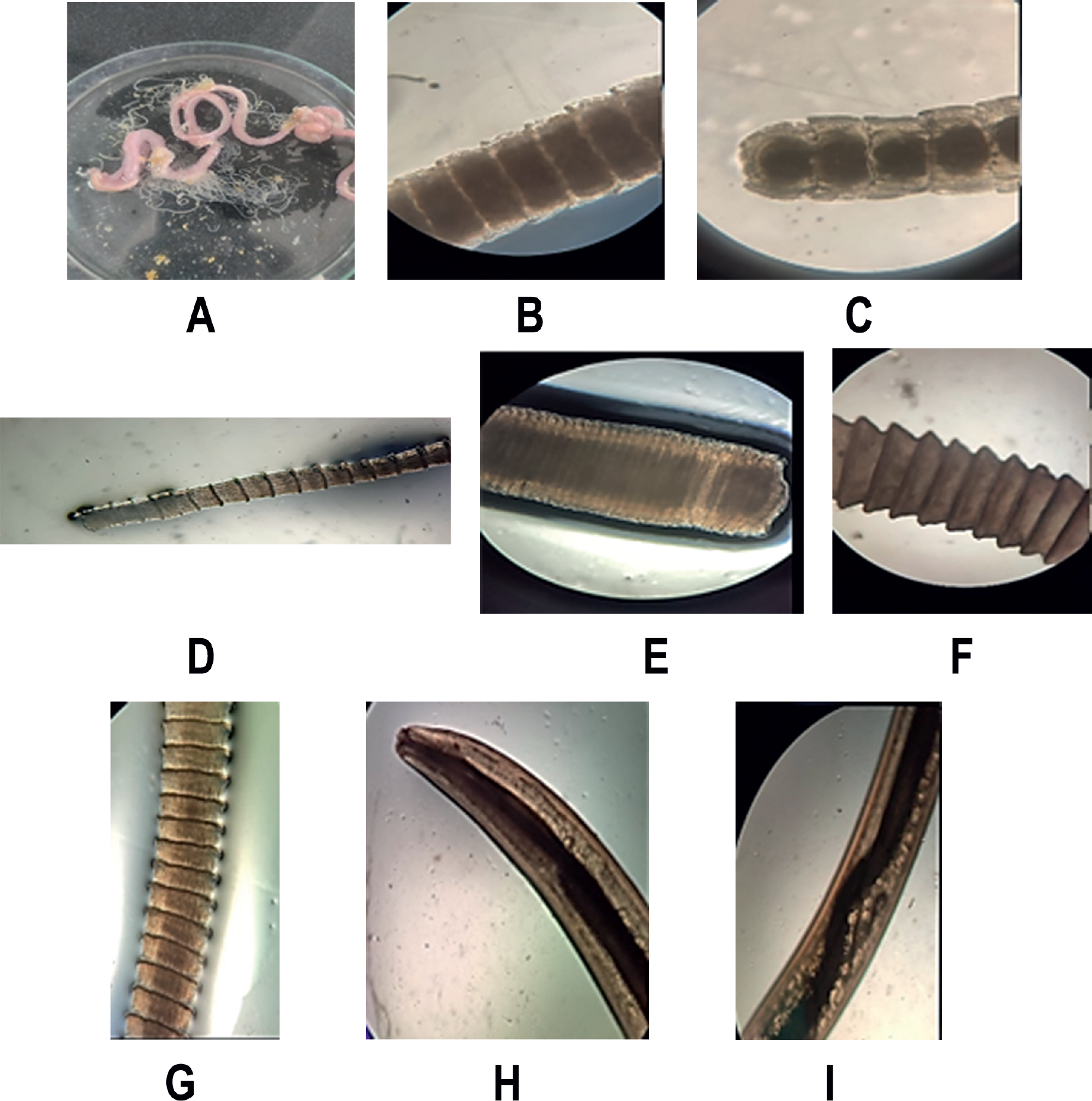
Fig 2.
The detected endoparasites. (A) The dissected intestine showing the released cestodes; (B) R. tetragona; (C) R. micracantha; (D) C. digonopora; (E) C. intermedia; (F) B. columba; (G) C. infundibulum; (H) A. columbae (anterior part); (I) A. columbae (middle region).
Table 3.
Demographic characteristics of the samples studied.
| Gender | Infected, N (%) | Uninfected, N (%) | P-value |
|---|---|---|---|
| Male (15) | 12 (80 | 3 (20) | 0.389 |
| Female (6) | 3 (50) | 3 (50) | |
| Total (21) | 15 (71.4) | 6 (28.5) |
Table 4.
Prevalence of nematode and cestode parasites infection, based on the sex of the 21 pigeons examined.
| Parasites | Male, N (%) | Female, N (%) | Total, N (%) | P-value |
|---|---|---|---|---|
| R. tetragona + R. micracantha | 9 (42.85) | 3 (14.28) | 12 (57.1) | 0.502 |
| C. digonopora | 5 (23.8) | 0 | 5 (23.8) | |
| C. intermedia | 1 (4.76) | 1 (4.76) | 2 (9.52) | |
| B. columbae | 1 (4.76) | 1 (4.76) | 2 (9.52) | |
| C. infundibulum | 2 (9.52) | 1 (4.76) | 3 (14.28) | |
| A. columbae | 3 (14.28) | 0 | 3 (14.28) |
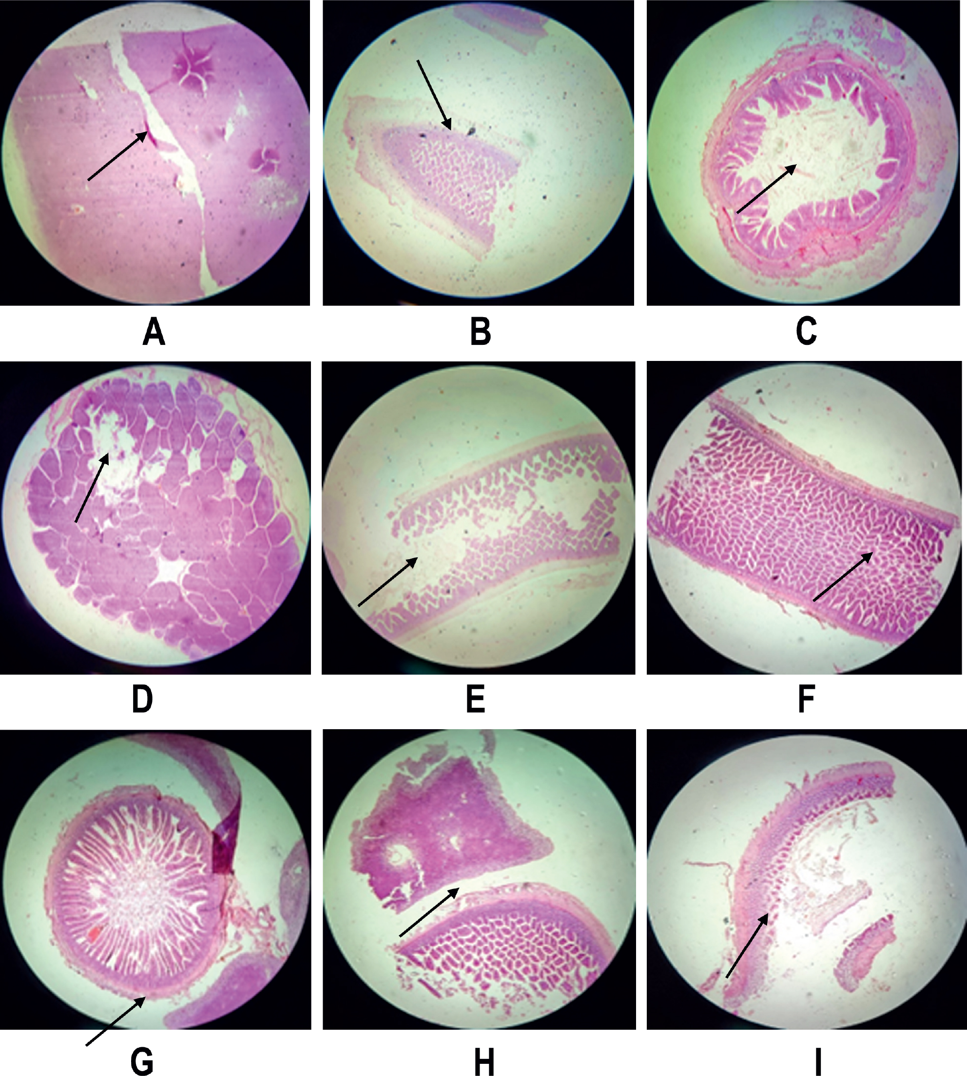
Fig 3.
Photomicrograph of intestine tissue sections (stained with H & E), at 40X. (A) Duodenal part infected with cestodes, shows destruction and shrinkage; (B) Atrophic hyperplasia in the infected ileum with cestode; (C) Rectum infected with cestodes shows inflammation of villi; (D) Inflammation in infected rectum with cestodes; (E) Inflammation in caecum infected with cestode; (F) Death of cells in infected intestine with nematode; (G) Migratory tunnels formation in intestine infected with nematode; (H, I) Intestine infected with nematode shows infiltration of lymphocytes, inflammation and histological hyperplasia.
Table 5.
Detected quadruple parasitic infection in relation to the pigeon sex.
| Male (N=1, 6.66%) | Female (N=1, 6.66%) | P-value |
|---|---|---|
| C. tschulyschma + R. tetragona + R. micracantha + C. intermedia | C. compar + R. tetragona + R. micracantha + B. columbae | 0.158 |
Table 6.
Detected triple cestode parasitic infection in relation to the pigeon sex.
| Parasites | Male, N (%) | Female, N (%) | Total, N (%) | P-value |
|---|---|---|---|---|
| R. tetragona + R. micracantha + C. digonopora | 5 (41.66) | 0 | 5 (41.6) | 0.375 |
| R. tetragona + R. micracantha + C. intermedia | 1 (8.33) | 1 (8.33) | 2 (16.6) | |
| R. tetragona + R. micracantha + B. columbae | 1 (8.33) | 1 (8.33) | 2 (16.6) | |
| R. tetragona + R. micracantha + C. infundibulum | 2 (16.66) | 1 (8.33) | 3 (24.9) | |
| Total | 9 (75) | 3 (25) | 12 (100) |