Helminthic infestation is a major health problem worldwide, more particularly in third world countries because of its prevalence there. Heavy rainfall, hot and humid climatic condition of Assam with vast areas of perennial ponds and ditches is congenial for propagation and multiplication of snail borne helminth parasites particularly the Paramphistomum cervi.
For several decades, the control of these parasitic infections has mainly relied on the repeated use of chemical anthelmintics provided by pharmaceutical companies (Charlier et al., 2022). But, from the field survey it was observed that rural farmers faced numerous constraints of using synthetic anthelmintics as it is not easily available in field and also, lack of knowledge on the proper use of them leads to toxicity of animals. This ultimately affect the production and worsen the economic condition of farmers. Again, environmental accumulation, anthelmintic resistance, and unreliable manufactures who at times unscrupulously produce anthelmintics with little or no efficacy at all become a serious issue. For these various reasons, interest in the screening of medicinal plants for their anthelmintic activity remains of great scientific interest despite extensive use of synthetic chemicals in modern clinical practices all over the world. Only screening of medicinal plants will not serve the purpose of using them as anthelmintics. Along with toxicity study of the plant, dose standardization as well as mechanism of action of the plant are equally important for development of an effective and safe alternative anthelmintics in near future.
Many medicinal plants are found in North East region of India but very less research have been undertaken to establish scientific proof for it. Zanthoxylum armatum DC is deciduous and evergreen shrub from the family Rutaceae. They are native to the World's warm temperate and sub-tropical regions. In India, Kashmir to Bhutan and also found in throughout North-East India. Prickly ash (English), Tejphal (Hindi), Tezowati (Sanskrit, Assamese and Bengali) and Timur (Nepal) are some of the common names for them. The different parts of the plants like leaves, roots, bark, seeds, fruits, stem, thorns are extensively used in the indigenous system of medicine for curing common illness like vomiting, diarrhoea, abdominal pain, cold, rheumatism, anthelmintic and traumatic injury etc. (Medhi et al., 2013). The fruits and seeds are used as piscicide, aromatic tonic in fever, dyspepsia, skin diseases and for expelling roundworms. Mehta et al., (2012) examined the methanolic seed extract of Z. armatum DC which induces worm paralysis (Pheretima posthuman) and death in a short period of time. Kumar et al., (2016) investigates the chemical composition and larvicidal activity of Z. armatum DC against diamondback moth, Plutella xylostella. Singh et al., (2016) studied the anthelmintic efficacy of aqueous extract of Z. armatum DC seeds against Haemonchus contortus.
Review of literature revealed the anthelmintic effect of Zanthoxyllym armatum DC but its mechanistic action as an anthelmintic is not yet established. Therefore, the present study was undertaken to evaluate anthelmintic effect of hydro-ethanolic seed extract of Zanthoxyllym armatum DC against P. cervi as well as its mechanistic evaluation for paralysis of worms.
The seeds of Zanthoxylum armatum DC was collected from Arunachal Pradesh and in and around Guwahati city, Assam, India. The samples were identified by Botanical Survey of India, Shillong, Meghalaya (File No. BSI/ERC/Tech/2022-23/1088). After collection, seeds were cleaned from any extraneous materials, then shade dried, powdered mechanically, weighed and stored in an airtight container till further use.
Exactly 250 g of powdered seed material was soaked in 700 ml of ethanol and 300 ml of distilled water (70:30) for 72 hrs in a glass beaker, the mixture was stirred every 24 hrs using a sterile glass rod. The filtrate was obtained after passing through muslin cloth and was concentrated in a rotary evaporator (EQUITRON, Roteva manufactured by MEDICA INSTRUMENT MFG. CO. Mumbai-400013) at 45–50°C under reduced pressure. The extract was further dried over a water bath at 37°C to get a semi-solid consistency. The extract was kept in an airtight container, labelled properly at 4°C until use.
Mature and healthy Paramphistomum cervi were collected from the rumen of freshly slaughtered cattle at local abattoir in warm (38 ± 1°C) Hanks Balanced Salt Solution (HBSS) in an insulated container and brought to the laboratory. The worms were kept in the BOD incubator at 38 ± 1°C until further use. The flukes were identified before experimentation using the procedure followed by (Kumar, 1999).
An acute oral toxicity study was performed according to the guidelines of Organization for Economic Co-operation and Development (OECD-423). The overnight fasted mice (n=3) were orally administered HESEZA at the limit dose of 2000 mg/kg body weight and observed continuously for behavioural, neurological and autonomic profiles for 30 min, 1hr, 2hr, 4hr, 6hr and then 24hr and 72hr and thereafter up to 14 days for any lethality, moribund state or death. The study showed no toxicity or moribund state for administration of HESEZA up to the end of the study period.
The hydro-ethanolic seed extract of Z. armatum DC was subjected to phytochemical screening as per the method described by (Harborne, 1973) for the presence of different phytoconstituents.
Mature and healthy P. cervi were collected from the rumen of freshly slaughtered cattle at local abattoir in warm (38 ± 1°C) Hanks Balanced Salt Solution (HBSS) in an insulated container and brought to the laboratory. The motile adult worms were cleaned with lukewarm normal saline and transferred in a beaker containing HBSS and were kept in an incubator at 38 ± 1°C until required for the experiment on the same day. For in vitro motility study, six adult P. cervi in each Petri dish having different dilutions of extracts (HESEZA) in HBSS ranging from 50, 150, 300, 500, 1000 and 3000 μg/ml and standard Oxyclozanide (10−5 M) were taken. However, control Petri dish received only HBSS. The total volume of HBSS in each Petri dish was kept at 5 ml. It was then incubated at 38 ± 1°C for 5 hrs and the number of live and dead adult worms were counted at 0, 30, 60, 90, 120, 150,180, 210, 240, 270 and 300 minutes of exposure as described by Sujon et al., (2008).
To elucidate the possible mechanism of action of the plant extract, various biochemical parameters were studied. The effect of HESEZA in various concentrations (100, 300, 1000 and 3000 μg/ml) and standard reference drug Oxyclozanide (10−5 M) (George, 2004) were studied. P. cervi (200 – 300 mg) were incubated in Petri dishes containing various concentrations of the plant extracts in HBSS for 4 hrs at 38 ± 1°C in an incubator. After incubation, the worms or the incubate were used for biochemical studies.
Glucose uptake study was conducted to detect the changes in glucose absorption by the rumen flukes in presence of plant extracts. These studies are based on the principle of (Van den Bossche, 1972; Ahmed & Nizami, 1987). After 4 hrs of incubation the amount of glucose left in the incubate was estimated by the method described by (Hultman, 1959). Aldohexoses present in deproteinized sample react with O-toluidine reagent to give blue green colour of N-glycosylamine. The intensity of colour developed in the test sample is compared with that of standard glucose solution at 630 nm (UV 2100, Chemito) and glucose content of the sample is computed.
After 4 hrs of incubation with extracts, the worms were washed 3 times with normal saline and their glycogen was precipitated and hydrolysed by method of (Hassid & Abraham, 1957). The hydrolysed glucose was estimated by the method described by (Hultman, 1959). The tissue containing glycogen was digested in hot concentrated KOH, precipitated with ethanol and then hydrolysed with acid. On hydrolysis, 1 g glycogen gives 1.11 g of glucose.
After 4 hrs of incubation, the worms were rinsed thrice with normal saline and their lactic acid was estimated by the method of (Gutmann & Wahlefeld, 1974). The lactate in protein free filtrate is converted to pyruvate by lactate dehydrogenase in presence of NAD. NADH formed in the reaction is measured by the increase in extinction at 340 nm (UV 2100, Chemito).
The activity of acetylcholinesterase of whole worms was estimated by the method described by (Fishman & Green, 1961). This method is based on the chemical determination of unreacted acetylcholine, which reacts with hydroxylamine to form acetyldroxamic acid, and further reacts with Fe+++ in acid solution to form a soluble red-purple colour complex, that can be estimated photometrically at 540 nm.
Faecal samples were screened for the presence of P. cervi infestation by sedimentation and floatation technique as per the standard method described by (Soulsby, 1982).
Faecal samples of affected goats were examined on on “0” day (pre-treatment), 7th day, 14th day, 21st day and 28th day post-treatment by using standard sedimentation and floatation technique. Determination of EPG of all the selected animals was carried out by the McMaster technique as described by Sloss et al., (1997) for five occasions at 7 days interval.
Blood samples collected with K1-EDTA anticoagulant were used for the determination of various haematological parameters i.e. Hb (g/dl), PCV (%), TEC (x106/mm3), TLC (x103/mm3), DLC: Neutrophil (%), eosinophil (%), basophils (%), lymphocytes (%) and monocytes (%) in all the groups on day 0, 7, 14, 21 and day 28 by using Automatic Haematology Analyser (MELET, SCHLOESING Laboratories, FLS257).
Various biochemical parameters such as serum total protein (g/dl), albumin (g/dl), globulin (g/dl), AST (U/L), ALT (U/L), ALP (U/L), serum iron (μg/dl), copper (μg/dl), sodium (mmol/L), potassium (mmol/L) and chloride (mmol/L) were estimated in all the groups on day 0, 7, 14, 21 and day 28 by using commercially available diagnostic kits with the help of semi-automated clinical chemistry analyser (BeneSpheraTMC61).
The animals with the highest parasitic load were selected for the present study. Twenty-four numbers of affected goats of almost similar body weight and age of either sex was divided randomly into four different groups containing six animals each. Group A considered as control without receiving any treatment, Group B and C were extract treated groups which received hydro-ethanolic seed extract of Zanthoxylum armatum DC @ 150 and 250 mg/kg b. wt. orally while Group D received standard reference drug oxyclozanide @ 15 mg/kg b. wt orally. Faecal samples of affected goats were examined. A booster dose of HESEZA @ 150 and 250 mg/kg b. wt. orally was administered at 21 days of post-treatment.
Data were analysed by using SPSS version 20.0 using one- and two-way ANOVA.
The study was conducted after approval from the Institutional Animal Ethics Committee (IAEC), AAU, Khanapara, vide approval No. 770/GO/Re/S/03/CPCSEA/FVSc/AAU/IAEC/21-22/941 dated 20.08.2022.
The percentage yield of the solid residue obtained during the extraction of HESEZA was found to be 4.96 % w/w respectively.
The result of qualitative phytochemical analysis of HESEZA is presented in Table 1, which revealed presence of alkaloid, terpenoid, diterpenes, flavonoid, steroid, tannin, glycoside, and phenolic compound.
Qualitative phytochemical analysis of HESEZA.
| SI. No | Active principle | Test applied | Result |
|---|---|---|---|
| 1. | Alkaloid | Hager's test | Positive |
| 2. | Terpenoids | Salkowski test | Positive |
| 3. | Diterpenes | Copper acetate test | Positive |
| 4. | Flavonoids | Ferric chloride test | Positive |
| 5. | Steroid | Salkowski test | Positive |
| 6. | Tannins | Ferric chloride test | Positive |
| 7. | Glycosides | Sodium hydroxide test | Positive |
| 8. | Saponins | Frothing test | Negative |
| 9. | Phenolic compound | Ferric chloride test | Positive |
The study revealed no lethality or toxic manifestation after oral administration of HESEZA up to the limit dose i.e 2000 mg/kg body weight in mice.
The gross motility of P. cervi was observed after incubating the worms at 38 ± 1° C for 5 hr of exposure in normal HBSS, different concentrations of seed extract and oxyclozanide (10−5 M). Reduction of motility of parasite was evident as feeble at 5 hr of exposure in presence of oxyclozanide (10−5 M). Similar type of result has also been observed with HESEZA @ 3000 μg/ml which is comparable with oxyclozanide @ 10−5 M. The results are presented in Table 2.
In vitro anthelmintic activity of different concentration of HESEZA against P. cervi.
| Drug/Extract | Conc (μg/ml) | No of parasite showing motility (in minutes) | ||||||||||
|---|---|---|---|---|---|---|---|---|---|---|---|---|
| 0 | 30 | 60 | 90 | 120 | 150 | 180 | 210 | 240 | 270 | 300 | ||
| Control | - | 6 | 6 | 6 | 6 | 6 | 6 | 6 | 6 | 6 | 6 | 6 |
| Z. armatum (Hydro-ethanolic seed extract) | 50 | 6 | 6 | 6 | 6 | 6 | 6 | 6 | 6 | 5 | 4 | 3 |
| 150 | 6 | 6 | 6 | 6 | 6 | 5 | 5 | 4 | 3 | 2 | 1 | |
| 300 | 6 | 6 | 6 | 5 | 4 | 3 | 1 | 0 | 0 | 0 | 0 | |
| 500 | 6 | 5 | 4 | 3 | 1 | 0 | 0 | 0 | 0 | 0 | 0 | |
| 1000 | 6 | 4 | 3 | 1 | 1 | 0 | 0 | 0 | 0 | 0 | 0 | |
| 3000 | 6 | 3 | 2 | 1 | 0 | 0 | 0 | 0 | 0 | 0 | 0 | |
| Oxyclozanide | 10−5 M | 6 | 2 | 0 | 0 | 0 | 0 | 0 | 0 | 0 | 0 | 0 |
Four important biochemical parameters viz., glucose uptake, glycogen content, lactic acid production and acetylcholinesterase activity affecting metabolism of the parasite i.e., P. cervi was undertaken.
The reference drug, oxyclozanide (10−5 M) produced significant changes in various biochemical parameters of incubated P. cervi in vitro. Glucose uptake was reduced significantly (P<0.05) to 1.11 ± 0.05 mg/100 mg of tissue, as compared to that of control (1.59 ± 0.09 mg/100 mg of tissue). Similarly, glucose uptake and glycogen content of P. cervi was reduced significantly (P<0.05) by HESEZA @ 300, 1000 and 3000 μg/ml on comparing with the control. The values attributes to glucose uptake parameter for HESEZA @ 300, 1000 and 3000 μg/ml are 1.34 ± 0.07, 1.19 ± 0.09 and 1.09 ± 0.08 mg/100 mg of tissue, on the other hand, the value of control is 1.52 ± 0.05 mg/100 mg of tissue. Again, the significant data extracted by HESEZA @ 300, 1000 and 3000 μg/ml for glycogen content parameter are 2.30 ± 0.08, 2.10 ± 0.03 and 1.87 ± 0.07 mg/100 mg of tissue, while for the control it is 2.54 ± 0.03 mg/100 mg of tissue. Again, lactic acid production was increased significantly (P<0.05) by the HESEZA @ 300, 1000 and 3000 μg/ml as compared to the control. The data obtained by the HESEZA @ 300, 1000 and 3000 μg/ml and control are 97.45 ± 6.40, 108.16 ± 4.18, 116.45 ± 3.27 and 80.47 ± 5.18. However, AChE activity of P. cervi was significantly (P<0.05) inhibited by HESEZA @ 3000 μg/ml (70.64 ± 3.66 mM of Ach hydrolyzed/g wet weight/h) as compared to control (92.09 ± 5.70 mM of Ach hydrolyzed/g wet weight/h). The results are presented in Table 3 and Figures 1, 2, 3 and 4.
In vitro effect of different concentration of HESEZA on various biochemical parameters following 4 hours of incubation of adult P. cervi.
| Biochemical parameters | Control | Standard (Oxyclozanide 10−3 M) | Concentrations (μg/ml) of seed extract | |||
|---|---|---|---|---|---|---|
| 100 | 300 | 1000 | 3000 | |||
| Glucose uptake (mg/100mg of tissue) | 1.52±0.05a | 1.08±0.03e | 1.45±0.07a | 1.34±0.07b | 1.19±0.09c | 1.09±0.08d |
| Glycogen content (mg/100mg of tissue) | 2.54±0.03a | 1.84±0.09e | 2.47±0.04a | 2.30±0.08b | 2.10±0.03c | 1.87±0.07d |
| Lactic acid production (μg/g) of tissue | 80.47±5.18a | 116.28±4.34e | 85.27±4.87a | 97.45±6.40b | 108.16±4.18c | 116.45±3.27e |
| Acetylcholinesterase activity (mM of Ach hydrolyzed/g weight/h) | 92.09±5.70a | 59.17±4.48e | 91.00±4.18a | 88.55±4.89ab | 83.00±4.19ab | 70.64±3.66c |
Values are the Mean ± S.E. for six replicates.
Mean value in each row bearing common superscripts do not differ significantly at P<0.05.
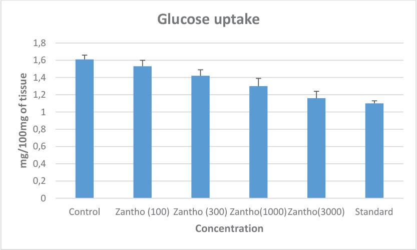
Effect of different concentration of HESEZA on glucose uptake in adult P. cervi.
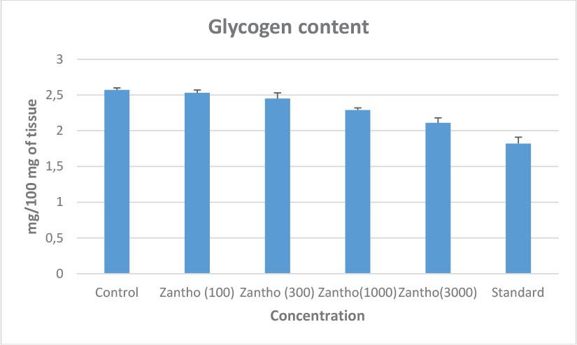
Effect of different concentration of HESEZA on glycogen content in adult P. cervi.
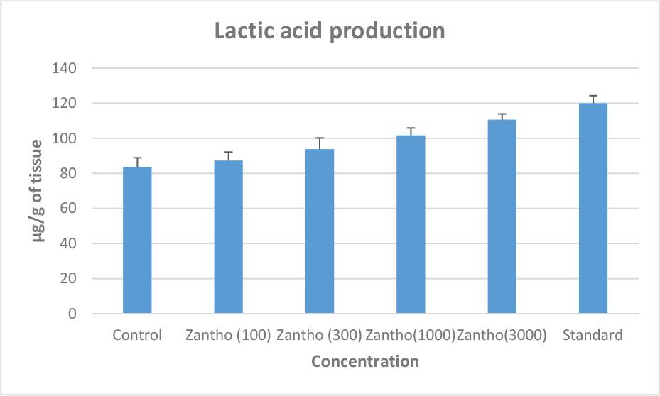
Effect of different concentration of HESEZA on lactic acid production in adult P. cervi.
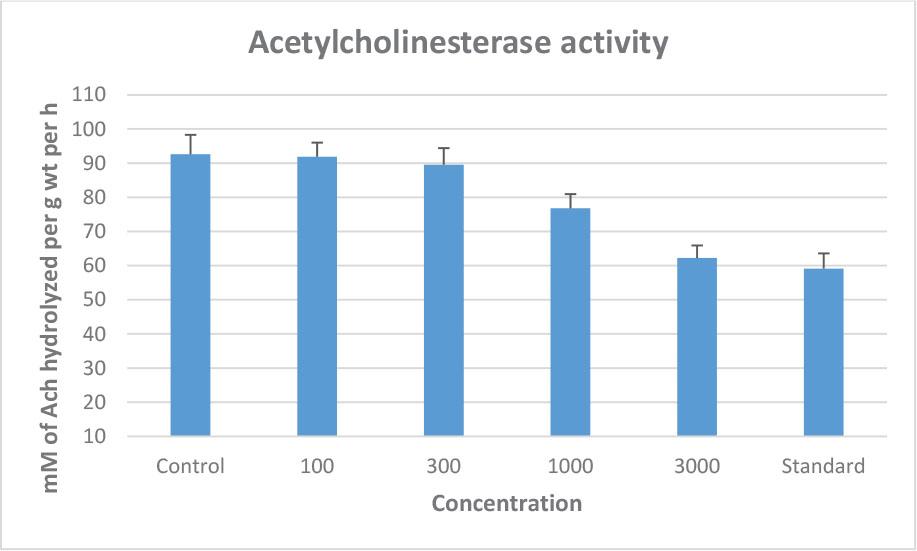
Effect of different concentration of HESEZA on acetylcholinesterase activity in adult P. cervi.
The mean ± S.E of EPG, in various groups are presented in Table 4. The efficacy for reduction of EPG for HESEZA @ 150 mg/kg b. wt at 7th, 14th, 21st and 28th days of post-treatment were 26.44, 48.98, 76.36 and 96.81 % while the same for 250 mg/kg b. wt were 35.48. 74.46, 92.37 and 100 % respectively. Again, the efficacy was maximum for the standard drug treated animals with 76.55, 98.44 at 7th, 14th day and 100 %, at 21st and 28th days of post-treatment.
In vivo study of HESEZA (150 and 250 mg/kg b. wt) on EPG in experimental groups at different days of treatment.
| Days of observation | |||||
|---|---|---|---|---|---|
| Group | 0th | 7th | 14th | 21st | 28th |
| A | 585±59.15a | 591.67±63.21a | 601.67±64a | 586.67±59.48a | 573.33±45.07a |
| B | 639±33.64a | 470±18.26ab | 326.33±12.33bc | 151.5±13.58c | 20.33±3.87d |
| C | 603.33±32.52a | 389.67±47.14b | 154.83±40.63c | 46.5±19.19d | 0±0 |
| D | 516.67±31.16a | 121.33±9.61b | 8±1.46c | 0±0 | 0±0 |
Values are the Mean ± S.E. for six replicates.
Mean value in each row bearing common superscripts do not differ significantly at P<0.05 and P<0.01.
Analysis of variance revealed that there was a highly significant difference (P<0.01) between different treatment groups, various days of treatment (P<0.01) and interaction between different treatment groups and various days of treatment (P<0.05).
The mean ± S.E of body weight (kg) in various groups are presented in Table 5. The body weight of extract treated animals and standard drug treated animals were increased significantly (P<0.05), although there was a non-significant difference between days of treatment.
In vivo study of HESEZA (150 and 250 mg/kg b. wt) on body weight in experimental groups at different days of treatment.
| Days of observation | |||||
|---|---|---|---|---|---|
| Group | 0th | 7th | 14th | 21st | 28th |
| A | 7.77±0.17a | 7.78±0.17a | 7.81±0.17a | 7.82±0.18a | 7.84±0.18a |
| B | 7.56±0.25a | 8.04±0.21ab | 8.61±0.23ab | 9.25±0.23b | 9.84±0.24b |
| C | 7.93±0.15a | 8.45±0.17ab | 9.11±0.2b | 9.85±0.21b | 10.88±0.26bc |
| D | 7.79±0.2a | 8.36±0.2ab | 9.32±0.3b | 10.26±0.19bc | 11.37±0.18c |
Values are the Mean ± S.E. for six replicates.
Mean value in each row bearing common superscripts do not differ significantly at P<0.05.
After treatment of infected animals with HESEZA and oxyclozanide, the hematological parameters especially Hb (g/dl), PCV (%), TEC (106/mm3), lymphocyte count (%) were increased significantly (P<0.05; P<0.01) in all the treatment groups. On the contrary, TLC (106/mm3), neutrophil (%), eosinophil (%) and monocyte count (%) were decreased significantly (P<0.05; P<0.01) in all the extract and standard drug treated groups. The results are presented in Table 6.
In vivo study of HESEZA (150 and 250 mg/kg b. wt) on hematological parameters in experimental groups at different days of treatment.
| Group | Parameter | Days of observation | |||||
|---|---|---|---|---|---|---|---|
| 0th | 7th | 14th | 21st | 28th | |||
| A | Hb (g/dl) | 7±0.06a | 6.97±0.06a | 6.97±0.04a | 6.9±0.07a | 6.85±0.08a | |
| B | 7.03±0.11a | 7.07±0.13a | 7.77±0.12ab | 8.02±0.12c | 8.28±0.11c | ||
| C | 7.1±0.1a | 7.17±0.08a | 7.32±0.08a | 7.83±0.1ab | 8.57±0.08c | ||
| D | 6.92±0.06a | 7.85±0.04ab | 8.05±0.04c | 8.6±0.04c | 9.25±0.08d | ||
| A | PCV (%) | 20.55±0.34a | 20.57±0.36a | 20.6±0.34a | 20.63±0.34a | 20.68±0.3a | |
| B | 21.85±0.28a | 21.88±0.29a | 21.97±0.3ab | 22.03±0.28b | 22.13±0.29b | ||
| C | 20.48±0.34a | 21.53±0.32a | 21.87±0.32a | 22.08±0.32b | 22.57±0.32b | ||
| D | 20.32±0.31a | 21.78±0.27a | 22.05±0.26b | 22.77±0.29b | 22.93±0.29b | ||
| A | TLC (103/mm3) | 15.78±0.4a | 15.86±0.43a | 16.24±0.21a | 16.17±0.2a | 16.09±0.29a | |
| B | 15.47±0.25a | 14.72±0.35ab | 13.68±0.31b | 12.33±0.31bc | 11.35±0.33c | ||
| C | 15.68±0.22a | 14.1±0.3ab | 12.63±0.35bc | 11.27±0.37c | 9.72±0.32d | ||
| D | 15.29±0.3a | 12.15±0.36bc | 10.8±0.27cd | 9.38±0.26d | 8.9±0.24e | ||
| A | TEC (106/mm3) | 10.48±0.17a | 10.28±0.27a | 10.34±0.2a | 10.41±0.22a | 10.38±0.19a | |
| B | 9.62±0.18a | 10.33±0.15a | 11.05±0.19ab | 11.8±0.19ab | 12.55±0.16b | ||
| C | 10.2±0.28a | 11.23±0.3ab | 12.07±0.29b | 13.02±0.28bc | 13.78±0.26c | ||
| D | 10.31±0.32a | 12.08±0.12b | 13.29±0.21bc | 13.7±0.16c | 13.77±0.19c | ||
| A | Neutrophil count (%) | 51.4±0.36a | 51.35±0.33a | 51.68±0.38 | 51.79±0.45 | 51.58±0.45 | |
| B | 52.63±1.03a | 48.74±0.67b | 44.33±0.83bc | 40.57±0.77c | 36.34±0.92d | ||
| C | 50.72±1.03a | 46.11±0.87b | 41.84±0.93c | 36.97±1.1d | 31.95±1.2e | ||
| D | 51.01±0.47a | 41.28±0.86b | 35.59±0.6c | 30.76±0.31d | 29.25±0.34e | ||
| A | Eosinophil count (%) | 5.62±0.21a | 5.63±0.2a | 5.66±0.2a | 5.69±0.2a | 5.7±0.22a | |
| B | 5.11±0.13a | 4.53±0.14b | 3.88±0.1c | 3.19±0.11c | 2.42±0.12d | ||
| C | 5.6±0.28a | 4.92±0.32b | 4.18±0.33b | 3.52±0.31c | 2.65±0.26d | ||
| D | 5.51±0.17a | 4.6±0.17b | 3.83±0.17c | 3.05±0.17c | 2.25±0.15d | ||
| A | Monocyte count (%) | 4.08±0.13a | 4.08±0.12a | 4.08±0.13a | 4.13±0.14a | 4.1±0.13a | |
| B | 4.17±0.09a | 3.92±0.09a | 3.55±0.14b | 3.24±0.14b | 2.9±0.11bc | ||
| C | 3.98±0.12a | 3.64±0.13ab | 3.24±0.11b | 2.88±0.1bc | 2.36±0.11c | ||
| D | 4.1±0.12a | 3.47±0.14b | 3.05±0.11bc | 2.62±0.16c | 2.29±0.14c | ||
| A | Lymphocyte count (%) | 35.17±1.35a | 35.04±1.14a | 35.41±1.29a | 35.05±1.35a | 35.21±1.28a | |
| B | 34.17±1.07a | 38.21±1.06b | 42.26±1.04c | 46.8±0.82cd | 51.37±0.63e | ||
| C | 35.62±0.93a | 40.11±0.93bc | 45.04±0.89c | 49.77±0.88d | 54.91±0.8e | ||
| D | 35.74±0.7a | 42.72±1.11bc | 48.92±1.16c | 55.24±1.39d | 60.8±1.27e | ||
Values are the Mean ± S.E. for six replicates.
Mean bearing different superscripts in column differ significantly (P<0.05; P<0.01) in groups.
The biochemical parameters namely total serum protein (g/dl), serum albumin (g/dl), serum globulin (g/dl), serum iron (μg/dl), serum copper (μg/dl), serum sodium level (mmol/L), serum potassium level (mmol/L) and serum chloride level (mmol/L) were decreased in infected animals with P. cervi, but soon after treatment with HESEZA and oxyclozanide these parameters found to increased significantly (P<0.05; P<0.01) and attained its normal range within 28th days of post-treatment. However, the enzymatic profile such as aspartate aminotransferase level (U/L), alanine aminotransferase level (U/L) and alkaline phosphatase (U/L) were decreased significantly (P<0.05; P<0.01) to its normal range after post treatment with HESEZA and oxyclozanide. The results are provided in Table 7.
In vivo study of HESEZA (150 and 250 mg/kg b. wt) on biochemical parameters in experimental groups at different days of treatment.
| Group | Parameter | Days of observation | ||||
|---|---|---|---|---|---|---|
| 0th | 7th | 14th | 21st | 28th | ||
| A | Total serum protein (g/dl) | 4.28±0.11a | 4.24±0.09a | 4.26±0.1a | 4.25±0.1a | 4.23±0.09a |
| B | 4.27±0.09a | 4.67±0.11a | 5.03±0.17ab | 5.37±0.17ab | 5.79±0.16b | |
| C | 4.31±0.09a | 4.77±0.07a | 5.21±0.13ab | 5.62±0.13b | 6.03±0.14bc | |
| D | 4.38±0.08a | 4.84±0.13a | 5.23±0.14ab | 5.65±0.14b | 6.19±0.1bc | |
| A | Serum albumin (g/dl) | 1.82±0.04a | 1.81±0.04a | 1.81±0.03a | 1.81±0.03a | 1.82±0.04a |
| B | 1.75±0.03a | 1.96±0.05a | 2.26±0.08ab | 2.64±0.08b | 2.94±0.06bc | |
| C | 1.83±0.03a | 2.21±0.08ab | 2.64±0.11b | 2.97±0.09bc | 3.24±0.09c | |
| D | 1.8±0.04a | 2.19±0.09ab | 2.64±0.08b | 2.99±0.08bc | 3.26±0.08c | |
| A | Serum globulin (g/dl) | 2.27±0.05a | 2.24±0.05a | 2.25±0.05a | 2.25±0.06a | 2.26±0.06a |
| B | 2.16±0.08a | 2.35±0.06ab | 2.56±0.07b | 2.77±0.06c | 2.94±0.05d | |
| C | 2.24±0.09a | 2.45±0.08ab | 2.66±0.07b | 2.84±0.06c | 3.03±0.06d | |
| D | 2.15±0.07a | 2.39±0.07ab | 2.59±0.08b | 2.85±0.06c | 3.06±0.06d | |
| A | Aspartate aminotransferase level (U/L) | 102.65±2.04a | 102.95±1.82a | 103.32±1.7a | 103.32±1.69a | 103.7±1.51a |
| B | 100.89±1.51a | 93.28±1.69ab | 85.82±1.56b | 78.27±1.64bc | 70.29±1.43c | |
| C | 102.99±1.46a | 92.93±1.24ab | 84.92±1.37b | 76.84±1.04bc | 67.62±1.14c | |
| D | 103.75±2.16a | 90.85±1.82ab | 82.07±1.95b | 71.54±1.22bc | 62.49±1.11cd | |
| A | Alanine aminotransferase level (U/L) | 27.81±0.6a | 27.8±0.61a | 28.21±0.57a | 28.22±0.53a | 28.39±0.57a |
| B | 26±0.7a | 24.85±0.62b | 23.86±0.55c | 21.9±0.39d | 20.54±0.44d | |
| C | 27.23±0.38a | 25.5±0.54b | 23.71±0.4c | 22.12±0.36cd | 19.83±0.6de | |
| D | 27.38±0.55a | 25.47±0.62b | 23.02±0.43c | 20.57±0.55d | 18.96±0.36e | |
| A | Alkaline phosphatase level (U/L) | 189.72±1.17a | 190.44±1.36a | 191.76±1.5a | 192.05±1.19a | 192.5±1.2a |
| B | 189.38±1.42a | 177.65±1.31ab | 167.06±1.69bc | 156.64±1.74cd | 145.1±1.47d | |
| C | 188.25±1.43a | 176.9±1.77ab | 164.18±2.28c | 152.66±1.84d | 140.79±2.2e | |
| D | 189.1±2.08a | 174.96±1.76b | 161.76±1.48c | 148.04±1.03d | 136.47±1.22e | |
| A | Serum iron (µg/dl) | 150.26±1.42a | 150.32±1.42a | 150.29±1.43a | 150.26±1.44a | 150.25±1.43a |
| B | 152.07±1.54a | 155.1±1.5ab | 159.68±1.63b | 163.59±1.6c | 167.52±1.37cd | |
| C | 152.52±1.3a | 158.22±1.67b | 163.05±1.63c | 168.7±1.75cd | 173.5±1.73d | |
| D | 152.2±1.1a | 157.53±1.15b | 162.54±1.17c | 167.55±0.86cd | 172.64±0.81d | |
| A | Serum copper (µg/dl) | 61.99±0.88a | 61.83±0.82a | 61.93±0.83a | 61.99±0.84a | 62.03±0.84a |
| B | 62.39±1.48a | 65.17±1.51ab | 68.11±1.46b | 71.48±1.16bc | 75.38±1.28c | |
| C | 62.63±1.5a | 66.94±1.2ab | 71.95±1.29bc | 76.16±1.31c | 80.61±1.05cd | |
| D | 60.88±1.38a | 66.86±1.13ab | 72.33±1.15bc | 77.77±0.85c | 83.54±0.8cd | |
| A | Serum sodium level (mmol/L) | 124.06±1.4a | 123.67±1.31a | 123.5±1.21a | 123.72±1.16a | 123.84±1.13a |
| B | 125.93±0.9a | 127.79±0.94ab | 129.5±0.95b | 131.5±0.97bc | 133.18±0.8bc | |
| C | 123.51±1.18a | 125.91±0.98a | 128.53±1.01ab | 131.09±0.94bc | 134.1±1.05c | |
| D | 124.01±1.31a | 128.83±1.38ab | 132.05±1.53bc | 135.62±1.58c | 139.14±1.52d | |
| A | Serum potassium level (mmol/L) | 3.86±0.15a | 3.84±0.15a | 3.85±0.15a | 3.86±0.15a | 3.87±0.15a |
| B | 3.22±0.17a | 4.37±0.15bc | 4.5±0.15c | 4.58±0.15cd | 4.69±0.15d | |
| C | 3.59±0.15a | 4.01±0.08ab | 4.24±0.07b | 4.52±0.09cd | 4.75±0.07d | |
| D | 3.92±0.09a | 4.29±0.13b | 4.5±0.11cd | 4.69±0.09d | 4.93±0.09e | |
| A | Serum chloride level (mmol/L) | 69.79±0.95a | 69.68±0.97a | 69.77±0.99a | 69.87±0.98a | 70.02±0.93a |
| B | 71.27±1.89a | 74.41±1.92b | 76.75±2.01bc | 80.27±2.24c | 84.92±2.34d | |
| C | 71.09±2.09a | 74.76±1.94b | 79.02±2.07c | 84.16±1.51d | 88.91±1.12e | |
| D | 69.37±1.64a | 74.95±1.69b | 81±1.78cd | 88.67±2.24e | 96.55±2.08f | |
Values are the Mean ± S.E. for six replicates.
Mean bearing different superscripts in column differ significantly (P<0.05; P<0.01) in groups.
Research on medicinal plants especially on pharmacological studies and clinical trials have been conducted to systematize and validate ethno-veterinary practices and folklore claims of herbal drugs with the goal to use them in animal treatment and production. The common pharmacological basis of treatment of helminths generally involves interference with one or more vital functions of the parasite viz.:
Disruption of energy generation process and subsequent starvation of the parasite.
A break in the neuromuscular coordination causing paralysis of the parasites and subsequent expulsion and
Adverse effect on reproductive process.
Parasitic organisms have to conserve a reservoir of energy to continue their mechanical activity, growth as well as for reproductive behaviour (Bandyopadhyay et al., 2022). The carbohydrate metabolism and the rate limiting enzymes of the parasite are the major targets of currently used anthelmintics. These targets include, inhibition of glucose uptake, impairment of glycolysis, disruption of glycogen metabolism, blocking of fumarate reductase pathway and uncoupling oxidative phosphorylation (Van dan Bossche, 1980; Wang, 1984; Sharma, 1987; McKellar & Kinabo, 1991). The adult parasitic helminth completely depends on carbohydrates for their energy metabolism in contrast to their mammalian host. Glucose is the only direct source of energy for parasitic trematodes (Tielens, 1997). Thus, hindrance in the uptake of glucose will affect the motility of the flukes. However, in the absence of readily available glucose, the parasite may mobilize its reserve glycogen to maintain its live support mechanism. Accumulation of glycogen is a marked feature of parasitic helminth (Bandyopadhyay et al., 2022). Most of the anthelmintics act either by inhibiting glucose uptake and depleting the glycogen reserve of the parasite (Martin, 1997). Consequently, the energy production of the parasite is blocked. Decline in energy causes inhibition of motility of the flukes. Again, the elevated tissue lactic acid level implies that either malate pathway is inhibited or excretion of lactic acid from the parasite is blocked (Rahman & Bryant, 1977; Pampori et al., 1984). Hindrance in the malate pathway causes the inhibition of ATP production. So, the anaerobic glycolysis will be favored in which lactic acid is the end product. Thus, lactic acid is accumulated in the parasite which could be deleterious to the fluke. Motility of the parasite is declined due to lack of ATP.
To elucidate the mechanism of action of HESEZA, four important biochemical parameters viz., glucose uptake, glycogen content, lactic acid production and acetylcholinesterase activity affecting metabolism of the parasite i.e., P. cervi was undertaken.
The reference drug, oxyclozanide (10−5 M) produced significant changes in various biochemical parameters of incubated P. cervi in vitro. Glucose uptake and glycogen content of the standard drug treated helminths were reduced significantly (P<0.05) as compared to that of control. Similar trend of results has been attributed to HESEZA @ 300, 1000 and 3000 μg/ml which was significant @ P<0.05 on comparing with the control. Again, significant (P<0.05) increase of lactic acid accumulation in the tissue of P. cervi was observed on incubating the helminths with HESEZA @ 300, 1000 and 3000 μg/ml of concentration which was comparable to the standard drug @ 10−5 M. However, AChE activity of P. cervi was significantly (P<0.05) inhibited by HESEZA only @ 3000 μg/ml. Therefore, from the biochemical experiment, it can be now interpreting that HESEZA pocesses potential anti-trematodal property affecting the energy generation process in helminths resulting death and paralysis of P. cervi.
From the in vivo experiment in goats with P. cervi, it has been observed that goats treated with HESEZA @ 250mg/kg b. wt. showed highest decline in EPG count till 28th day post treatment. However, standard drug treated group (Oxyclozenide @ 15mg/kg b. wt) showed the best activity.
Although the effectiveness of HESEZA against trematode especially P. cervi is novel but some of the works has already been conducted in respect of seed extract of Z. armatum DC against earthworm and nematodes. Mehta et al., (2012) examined the methanolic seed extract of Z. armatum which induces worm paralysis and death of Pheretima posthuma (earthworm). Likewise, Singh et al., (2016) studied the anthelmintic efficacy of aqueous extract of Z. armatum DC seeds against Haemonchus contortus. The in vivo study of hydroethanolic seed extract of Zanthoxyllum armatum individually or in combination with methanolic seed extract of Entada phaseoloides and ethanolic seed extract of Butea frondosa were effective against gastrointestinal parasites of goats (Hussain, 2022). Now to discuss the anthelmintic efficacy of HESEZA, it is believed phytoconstituents mainly tannins, phenolic compounds and flavonoids present in seed extract are important and it can be supported by the study of Singh and Singh, (2011) who recorded different phytoconstituents in Z. armatum, responsible for the anthelmintic activity of the plant.
In the present study, a significant (P<0.05) increase in body weight was observed in all the treatment groups when compared with the control group. The increase in body weight might be due to the improved appetite of the animal after elimination of the flukes which was evidenced by the reduction in EPG in treated animals.
The hematological findings of the present study are similar to earlier reports explored by Bordoloi et al., (2012), Moudgil et al., (2017) and Shashank et al., (2019). The lowering of these parameters in infected goats might be due to acute loss of blood by sucking activity with resultant hemorrhages caused by parasites (Amulya et al., 2014). Since after post-treatment, the parasitic load was reduced drastically, thereby improving the blood profile of the infected animals.
Again, treatment of infected goats with HESEZA revealed improvement of biochemical parameters especially total serum protein, serum albumin, and all the essential micronutrient such as iron, copper, sodium, potassium and chloride which were decreased from normal range during the heavy infection with P. cervi in goats. Likewise, the enzymes namely aspartate aminotransferase, ala-nine aminotransferase, alkaline phosphatase that envisage the hepatic health was above the normal but during post treatment with the seed extract and oxyclozanide the enzymes came to a normal level. Similar type of findings was also reported by Hassan et al., (2012), Qamar and Maqbool, (2012), Ahmed et al., (2015), and Shashank et al., (2019) suggesting that decreasing of protein and all the essential micronutrients in the affected animals could be attributed to protein losing gastroenteropathy in fluke infestation and malabsorption of nutrient from damaged intestinal mucosa in concurrent gastrointestinal infections. Moreover, poor appetite due to hepatic loss during infection also synergize malnutrition of the animals. Soon after treatment with HESEZA, all the biochemical parameters came to a normal range as because the extract pocesses one most important phytoconstituent called flavonoid which might have improved the damaged liver and thereby improve the appetite and malnutrition as well.
The results of this study helped to establish the mechanism of action of seeds extract of Z. armatum DC against P. cervi which is an important objective in traditional medicine. From the above study, it is now considered that HESEZA affect the energy generation process of P. cervi causing death and paralysis of worms. These findings may help further for the development of alternative anti-trematodal medications without any resistance creation. Again, isolation and standardization of pure active phytoconstituent especially tannins, phenolic compound and flavonoid will be required for future experiment during the journey of effective alternative drug development.