
Table 1.
Genetic distances between Heterorhabditis kaankabi n. sp. and the Heterorhabditis species in the 28S and ITS ribosomal gene sequences.
| Species | Heterorhabditis kankabi n. sp. | |||
|---|---|---|---|---|
| 28S | ITS | |||
| p | N | p | N | |
| Heterorhabditis taysearae | na | na | 1.22 | 11 |
| Heterorhabditis mexicana | 0.67 | 6 | 1.22 | 11 |
| Heterorhabditis floridensis | 0.67 | 6 | 0.66 | 6 |
| Heterorhabditis baujardi | 0.67 | 6 | 1.37 | 11 |
| Heterorhabditis amazonensis | 0.79 | 7 | 1.66 | 15 |
| Heterorhabditis noenieputensis | 2.92 | 26 | 9.43 | 83 |
| Heterorhabditis indica | 2.92 | 27 | 9.01 | 79 |
| Heterorhabditis zealandica | 5.77 | 51 | 24.66 | 215 |
| Heterorhabditis megidis | 6.00 | 53 | 22.45 | 194 |
| Heterorhabditis downesi | na | na | 21.69 | 187 |
| Heterorhabditis safricana | 5.20 | 46 | 22.03 | 191 |
| Heterorhabditis marelatus | 5.20 | 46 | 21.91 | 190 |
| Heterorhabditis atacamensis | 4.88 | 42 | 20.65 | 179 |
| Heterorhabditis bacteriophora | 4.68 | 42 | 21.81 | 193 |
| Heterorhabditis georgiana | 5.10 | 45 | 22.03 | 195 |
| Heterorhabditis beicherriana | 4.95 | 43 | 22.03 | 195 |
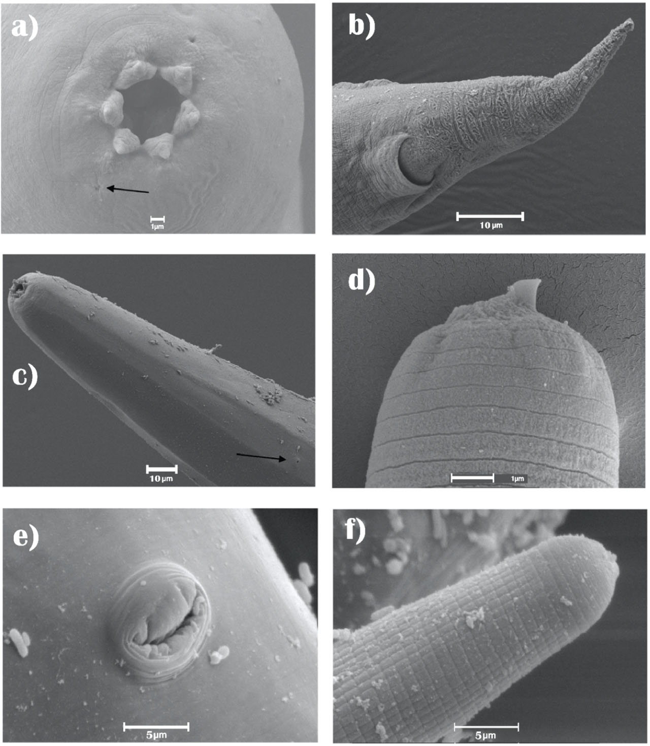
Fig. 1.
Heterorhabditis kankabi n. sp. SEM microphotographs. A, Head of an amfimictic female with six labial papillae and two amphids (magnification 5,000×); B, Posterior region of a hermaphrodite nematode showing anal area and tip of the tail (magnification 2,500×); C, Anterior region of the nematode where the excretory pore is observed in a hermaphroditic specimen (magnification 1,000×); D, infective juvenile with larval tooth and striated cuticle of the second instar (magnification 5,000×); E, Protruding vulvar region located in the middle part of the hermaphrodite specimen (magnification 5,000×); F, Anterior region of an ensheathed nematode showing the body with tessellate pattern (magnification 5,000×).
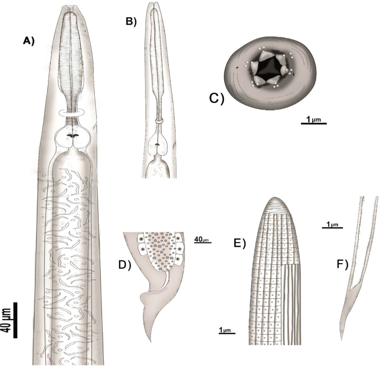
Fig. 2.
Schematic details of Heterorhabditis kankabi n. sp. A) anterior region of a hermaphrodite female; B) anterior region of an amphimictic female; C) apical view of a hermaphrodite female; D) tail of a hermaphrodite female; E) anterior region of an infective juvenile; F) tail of infective juvenile.
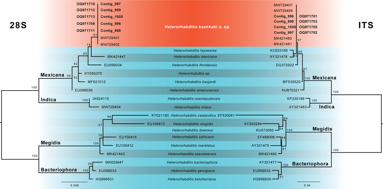
Fig. 3.
Phylogenetic relationships of species of the genus Heterorhabditis using the maximum likelihood method to generate the phylogenetic trees.
The tree shows ITS on the right side and the 28S on the left side.
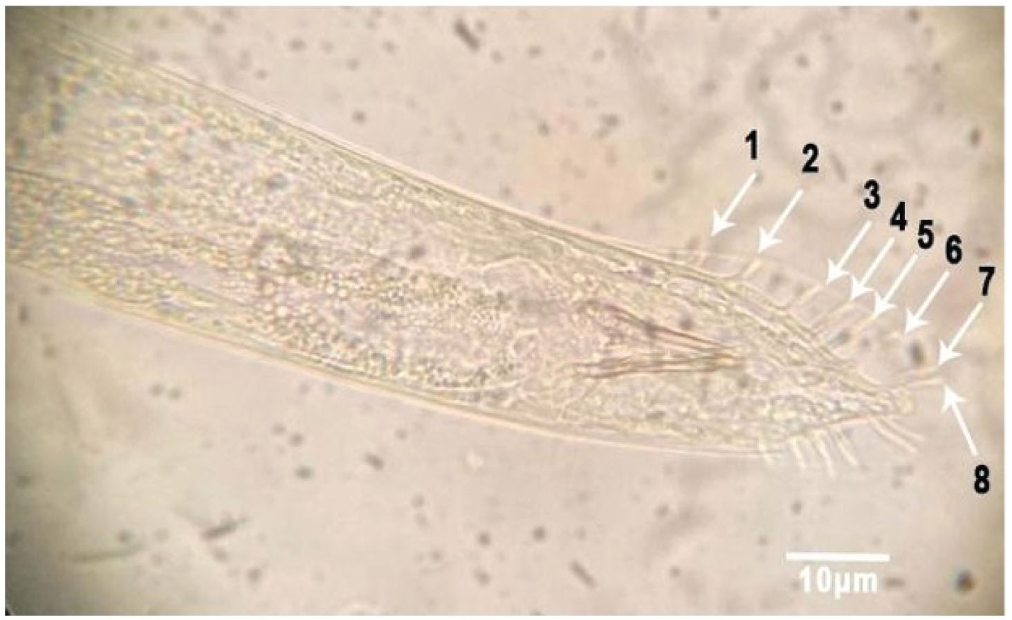
Fig. 4.
Clear field micrography of Metarhabditis rainai male ventral view showing eight bursal papillae.
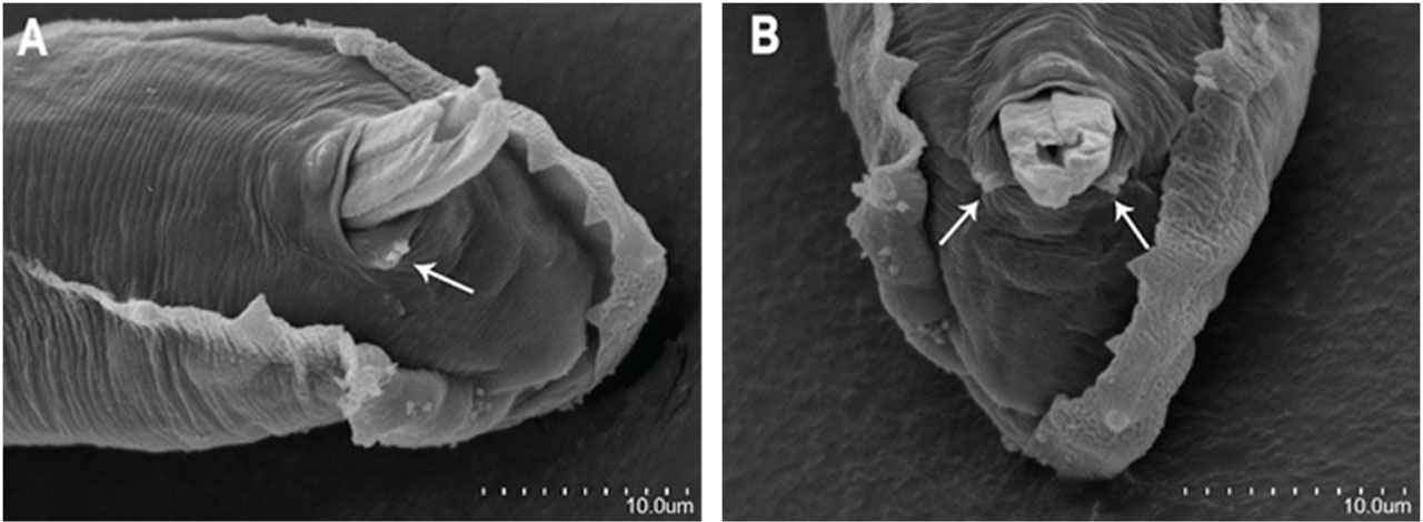
Fig. 5.
Metarhabditis rainai SEM microphotographs of male. A, posterior region lateral view bursal papillae with small adcloacal papillae, B, ventral view showing two pairs of small papillae on posterior edge of cloaca and spicules.
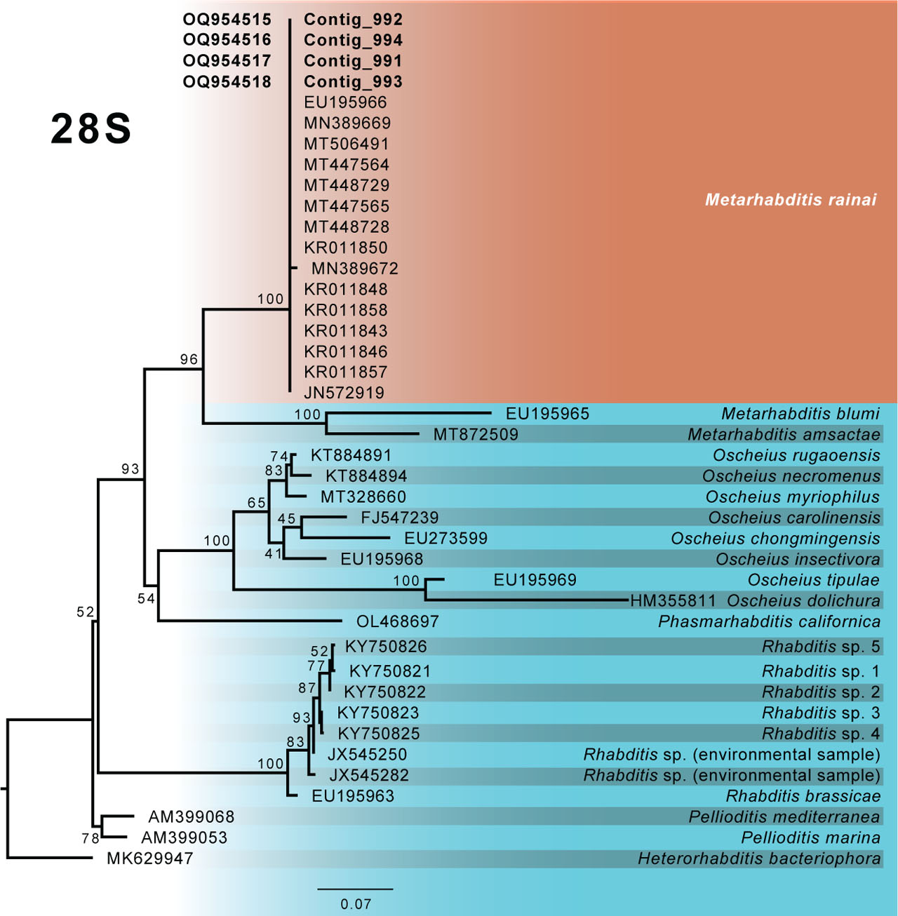
Fig. 6.
Phylogenetic relationships of species of the genus Metarhabditis using the maximum likelihood method to generate the phylogenetic trees.