
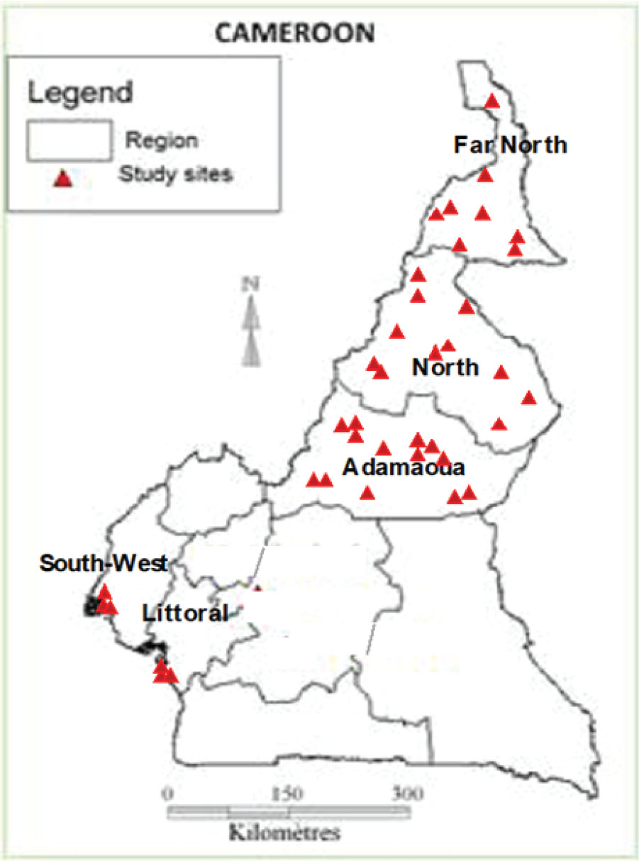
Fig. 1.
Map of schools investigated for soil-transmitted helminths (STH) infections.
Table 1.
Detailed morphological characteristics of filariform (L3) larvae of hookworm (Yoshida, 1966; WHO, 1981).
| Morphological Keys | A. duodenale | N. americanus |
|---|---|---|
| Length | 660 microns | 590 microns |
| Mouth | Less visible | 660 microns; Appears dark |
| Sheath | 720 microns; Less striated | Visibly striated and more clearly seen around the end of the body |
| Oesophagus and intestine | ¼ body length; no gap between the oesophagus and the intestine | ¼ body length; gap between the oesophagus and the intestine |
| Intestine | The anterior end is narrower in diameter than the oesophageal bulb | The anterior end is as wide as the oesophageal bulb |
| Tail | Blunt | Sharply pointed |
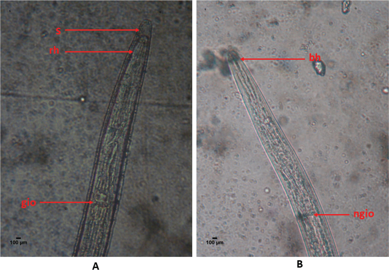
Fig. 2.
Head of a hookworm L3 larvae recovered from a hookworm-positive faecal sample
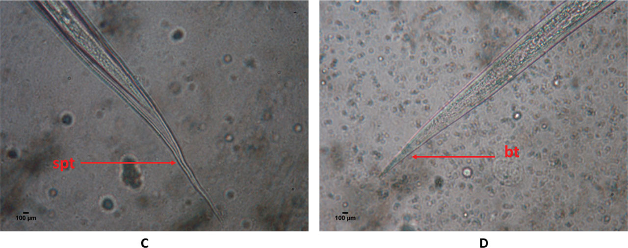
Fig. 3.
Tail of a hookworm L3 larvae recovered from a hookworm-positive faecal sample.
Table 2.
Number of L3 larvae characterized for hookworm species identification in five regions of Cameroon.
| N. americanus | A. duodenale | ||||
|---|---|---|---|---|---|
| Adamawa | 42 | 60 | 68.33 | 2852 | 0 |
| North | 75 | 10 | 40 | 315 | 0 |
| Far-North | 140 | 25 | 32 | 686 | 0 |
| South-West | 12 | 45 | 71.11 | 3565 | 0 |
| Littoral | 6 | 17 | 76.47 | 413 | 230 |
| Total | 275 | 157 | 57.58 | 7831 | 230 |
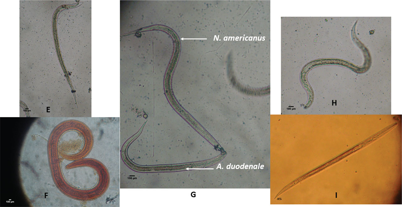
Fig. 4.
Hookworm entire L3 larvae recovered from hookworm-positive faecal samples.