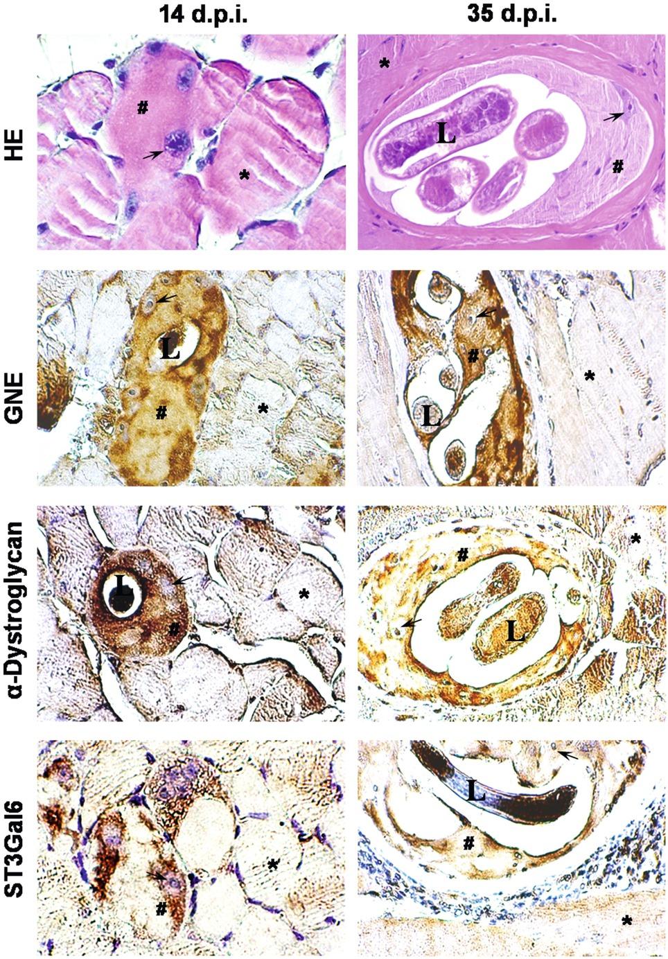
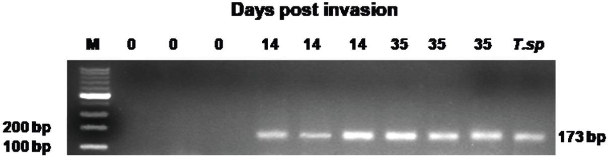
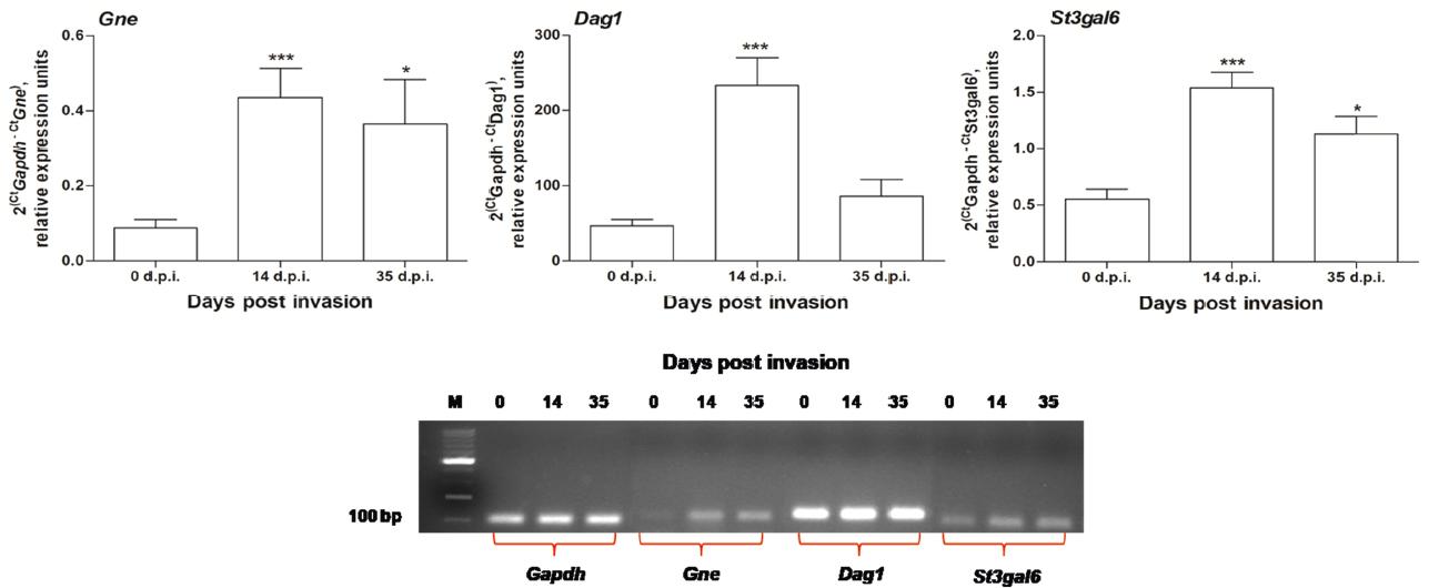
Have a personal or library account? Click to login
The synthesis of UDP-N-acetylglucosamine 2-epimerase/N-acetylmannosamine kinase (GNE), α-dystroglycan, and β-galactoside α-2,3-sialyltransferase 6 (ST3Gal6) by skeletal muscle cell as a response to infection with Trichinella spiralis Cover

The synthesis of UDP-N-acetylglucosamine 2-epimerase/N-acetylmannosamine kinase (GNE), α-dystroglycan, and β-galactoside α-2,3-sialyltransferase 6 (ST3Gal6) by skeletal muscle cell as a response to infection with Trichinella spiralis

Open Access
|Dec 2022

Figures & Tables

Fig. 1

Immunohistochemistry. Modified methacarn fixed sections from mouse skeletal muscles with Trichinella spiralis at days 14 and 35 post invasion (d.p.i.) were stained with rabbit polyclonal antibodies against glucosamine (UDP-N-acetyl)-2-epimerase/N-acetylmannosamine kinase (GNE), α-dystroglycan and ST3 beta-galactoside alpha-2,3-sialyltransferase 6 (ST3Gal6). Paralel sections were subjected to H&E staining to facilitate the histological orientation. Strong expressions of GNE and α-dystroglycan, and moderate expression of ST3Gal6 were observed on days 14 and 35 after invasion, suggesting these proteins as permanent characteristics of the Nurse cell of T. spiralis. The brown colour indicates positive immunohistochemical reaction, hashtag indicates the occupied sarcoplasm, star – non-occupied skeletal muscle cell, arrow – enlarged nucleus, L– larva. H&E, HRP anti-rabbit IgG, DAB. Scale bar 20 μm.

Fig. 2

Agarose gel analysis of Trichinella spiralis ESV fragment PCR. Polymerase chain reaction was performed on modified methacarn fixed mouse skeletal muscle tissue sections, selected on days 0, 14 and 35 after T. spiralis invasion. Genomic DNA from T. spiralis infectious larvae was used as a positive control sample. Presence of 173 bp fragment of expansion segment V of the T. spiralis genome was detected only in the mouse samples collected on days 14 and 35 after invasion. The photograph is a representative of three randomly selected samples from each experimental group.

Fig. 3

Expressions of mouse glucosamine (UDP-N-acetyl)-2-epimerase/N-acetylmannosamine kinase (Gne), dystroglycan 1 (Dag1) and ST3 beta-galactoside alpha-2,3-sialyltransferase 6 (St3gal6) analysed by real time RT-PCR in modified methacarn fixed mouse skeletal muscle tissue sections, selected on days 0, 14 and 35 after T. spiralis invasion. The graphs show the relative quantification of the gene expressions calculated by the ΔΔCt method versus glyceraldehyde phosphate dehydrogenase (Gapdh) as a reference gene from five individual samples in triplicate. The bars show the standard error of mean. The products of amplification were loaded on 2.5% agarose gel versus Perfect 100-1000 bp DNA Ladder.

The full names of the investigated genes and their primers sequences used in this study_

GeneAbbreviationSpeciesAccession numberPrimers sequences (5`-3`)Product size (bp)
Glyceraldehyde 3-phosphate dehydrogenaseGapdhMus musculusNM_001289726, transcript variant 1TCCTCGTCCCGTAGACAAAATG –F AATCTCCACTTTGCCACTGC – R103
Glucosamine (UDP-N- acetyl) – 2 – epimerase/N- acetylmannosamine kinaseGneMus musculusNM_015828.3AATCCTGCAGATGTGTGTGG –F AATGCAGCACAACTCCTTCC – R119
Dystroglycan 1Dag1Mus musculusNM_001276485.1, transcript variant 5GTTGGCATTCCAGACGGTAC –F AGTGTAGCCAAGACGGTAAGG – R136
ST3 beta-galactoside alpha- 2,3-sialyltransferase 6St3gal6Mus musculusNM_018784.2TCCCAGCTGAAGAAATGAGGAC –F TCAGCTCTGCACAGAAATGG – R112
Expansion segment VESVTrichinella spiralis*GTTCCATGTGAACAGCAGT –F CGAAAACATACGACAACTGC – R173
DOI: https://doi.org/10.2478/helm-2022-0027 | Journal eISSN: 1336-9083 | Journal ISSN: 0440-6605
Language: English
Page range: 217 - 225
Submitted on: Mar 22, 2022
|
Accepted on: Aug 17, 2022
|
Published on: Dec 17, 2022
In partnership with: Paradigm Publishing Services
Publication frequency: 4 issues per year

© 2022 R. Milcheva, K. Todorova, A. Georgieva, S. Petkova, published by Slovak Academy of Sciences, Institute of Parasitology
This work is licensed under the Creative Commons Attribution-NonCommercial-NoDerivatives 4.0 License.