
Fig. 1
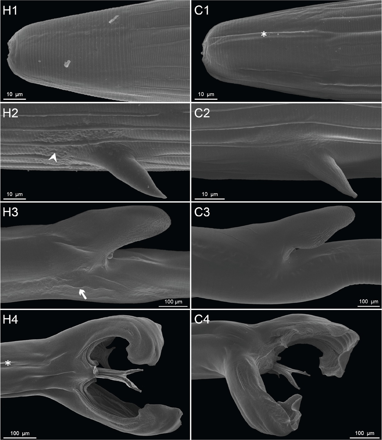
Scanning electron photomicrographs of Haemonchus contortus from the in vivo experimental groups. H1–H4 depict specimens collected from lambs fed a diet enriched with a Herbmix extract, and C1–C4 depict specimens from the control animals. H1 and C1, cephalic end of the body, both females; H2 and C2, region near the cervical papillae, both females; H3 and C3, vulvar region of the body, both females; H4 and C4, posterior end with the copulatory bursa, both males. Note the cephalic end of the female body (C1) and the posterior part of the male body (H4), with several artificial folds (asterisk), small protuberances (arrowhead) in the region anterior to the base of a cervical papilla (H2) and weak ridges (arrow) on the cuticle in the vulvar region (H3).
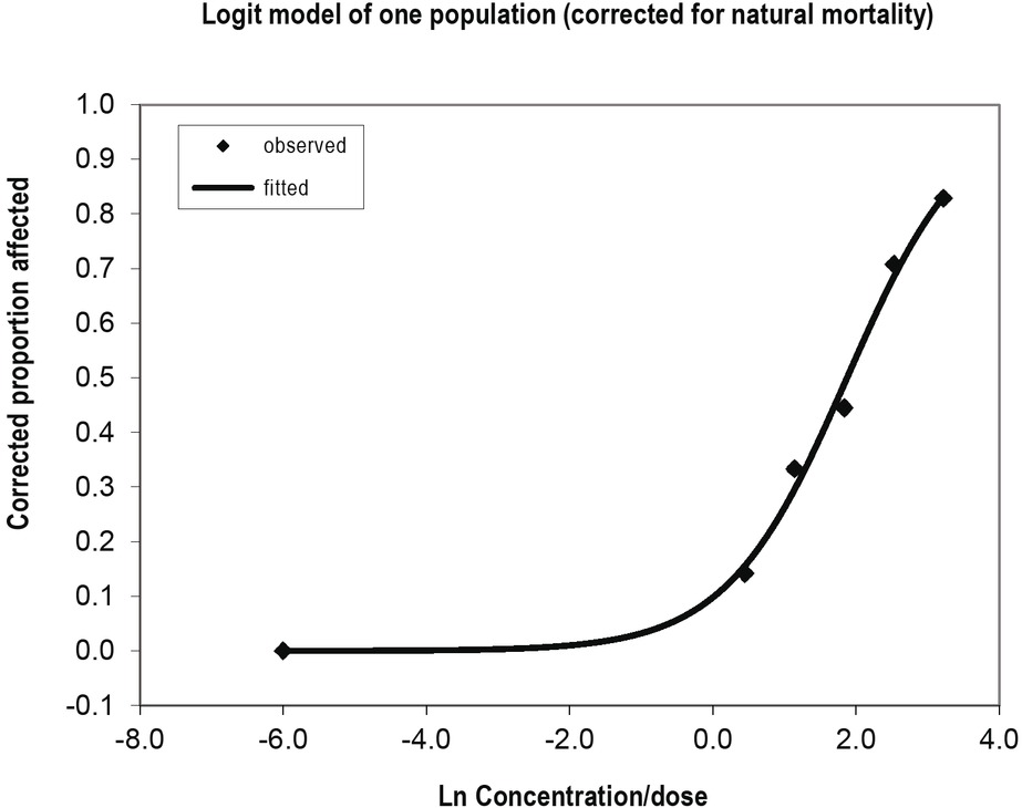
Fig. 2
Dose-response relationship of the Herbmix aqueous extract against Haemonchus contortus in the EHT.
Table 1
Hatching and ovicidal activity at the concentrations of the Herbmix aqueous extract tested in the EHT.
| Herbmix concentration (mg/mL) | Ovicidal effect (%) | Hatching (%) | p value |
|---|---|---|---|
| 25 | 83 | 17 | < 0.001 |
| 12.5 | 71 | 29 | < 0.001 |
| 6.25 | 45 | 54 | < 0.001 |
| 3.125 | 34 | 66 | < 0.001 |
| 1.563 | 15 | 85 | < 0.001 |
| Control | 1 | 99 | - |
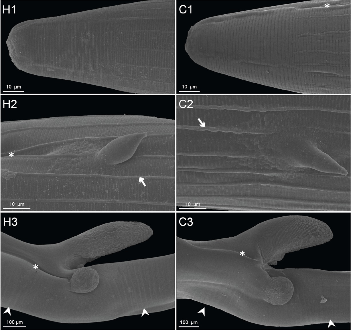
Fig. 3
Scanning electron photomicrographs of Haemonchus contortus from the in vitro experimental groups. H1–H3 depict specimens treated with the Herbmix extract diluted in PBS, and C1–C3 depict specimens from the control group kept in PBS. H1 and C1, cephalic end of the body; H2 and C2, region around a cervical papilla; H3 and C3, vulvar region with a lateral button. Note the slightly sinuous physiological longitudinal ridges (arrows), weak transverse wrinkles (arrowheads) and artifacts of sample preparation (asterisks).