
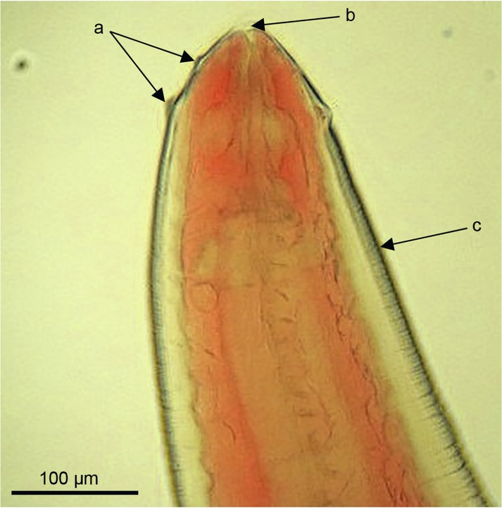
Fig. 1
The head end of Eustrongylides excisus larvae (L4). Visualised two rows of papillae (a) around the oral opening (b). The body of the larva is visibly striated (c) (Goncharov et al., 2018)
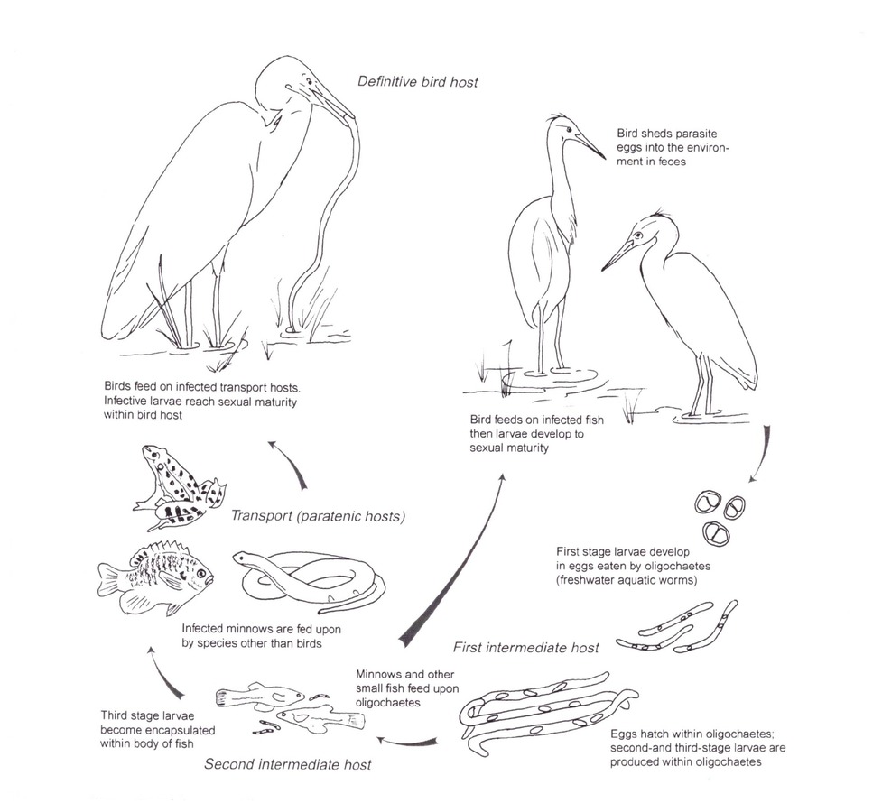
Fig. 2
Life cycle of Eustrongylides sp. (Cole, 1999)
Table 1
Human cases of eustrongylidosis infection.
| Country, area, year | Researched species | Human cases | Site of infection |
|---|---|---|---|
| USA, California, 1979 (Beaver & Thei, 1979) | Eustrongylides spp. | suspected Eustrongylides | Thorax |
| USA, Maryland, 1982 (Guerin et al., 1982) | Eustrongylides spp. | 2 cases | Abdominal cavity, intestine |
| USA, New Jersey, 1989 (Eberhard et al., 1989) | Eustrongylides spp. | 1 case | Abdominal cavity, intestine |
| USA, New York, 1989 (Wittner et al., 1989) | Eustrongylides spp. | 1 case | Abdominal cavity |
| USA,New Jersey, Big Timber Creek of Belmawr, 1996 (Narr et al., 1996) | E. ignotus | 1 case (17-year-old boy) | Abdominal cavity, intestine |
| South Sudan The Payam District of Jie, Kapoeta East County, Eastern Equatoria State, 2014 (Eberhard & Ruiz-Tiben, 2014) | Eustrongylides spp. | 2 cases | Distal segment of lower limbs |