
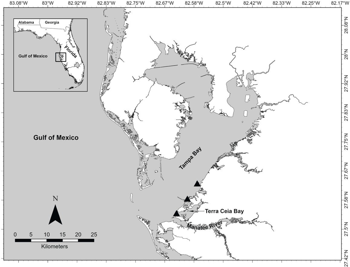
Fig 1
Map of Tampa Bay, Florida, USA, showing three collection localities for hosts, Tylosurus crocodilus, found to be infected by Philometra consimilis n. sp. or both that species and Barracudia notabilis n. sp.
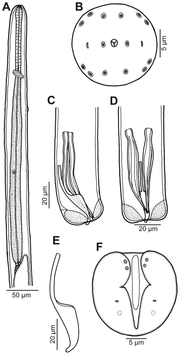
Fig. 2
Barracudia notabilis n. sp. from T. crocodilus, male. A – anterior end of body, lateral view; B – cephalic end, apical view; C, D – caudal end, lateral and ventral views, respectively; E – gubernaculum, lateral view; F – caudal end, apical view
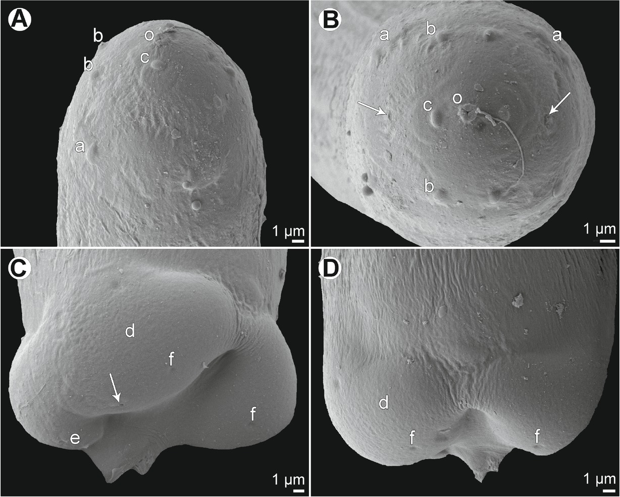
Fig. 3
Barracudia notabilis n. sp. from T. crocodilus, scanning electron micrographs of male. A, B – cephalic end, lateral and apical views, respectively (arrows indicate amphids); C, D – caudal end, sublateral and dorsal views, respectively (arrow indicates phasmid). Abbreviations: a – pair of cephalic papillae of outer circle; b – submedian cephalic papilla of inner circle; c – lateral cephalic papilla of inner circle; d – caudal mound; e – two adanal papillae; f – cuticular depression; o – oral aperture
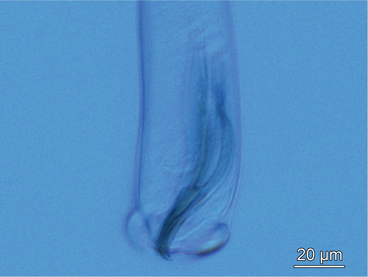
Fig. 4
Caudal end of Barracudia notabilis n. sp., lateral view
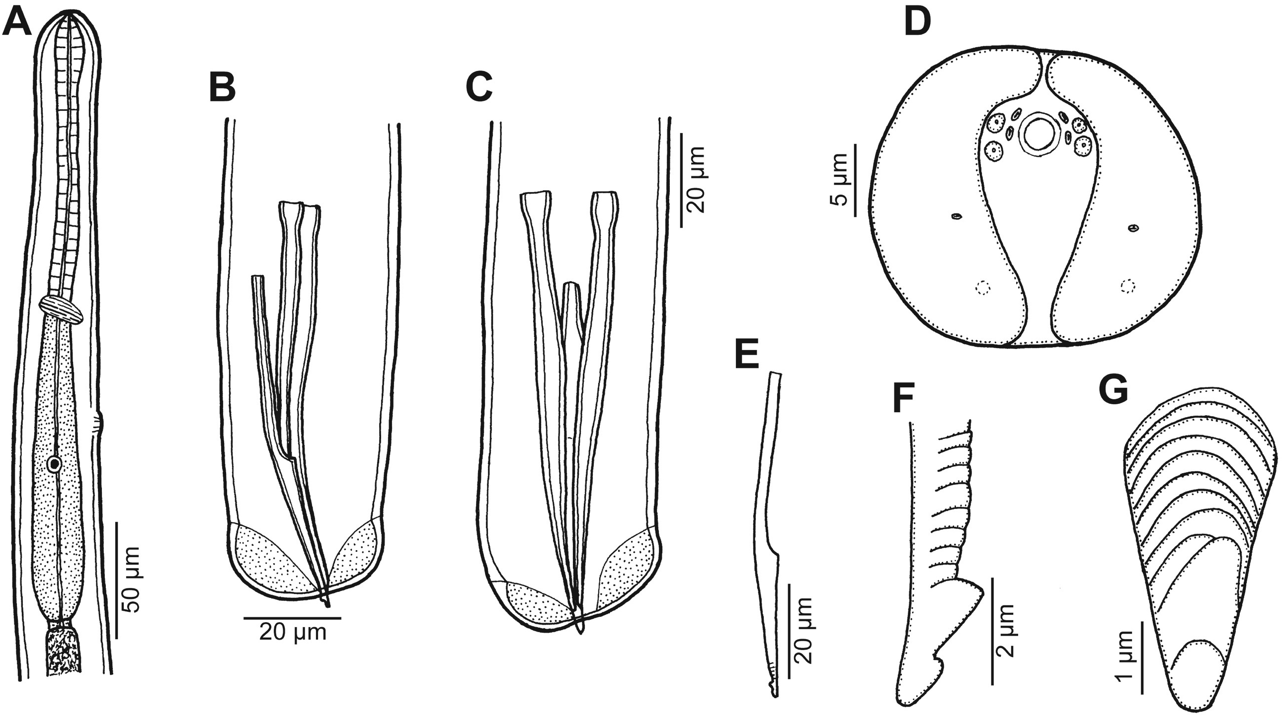
Fig. 5
Philometra consimilis n. sp. from T. crocodilus, male. A – anterior end of male, lateral view; B, C – posterior end of male, lateral and ventral views, respectively; D – caudal end, apical view; E – gubernaculum, lateral view; F, G – distal tip of gubernaculum, lateral and subdorsal views, respectively
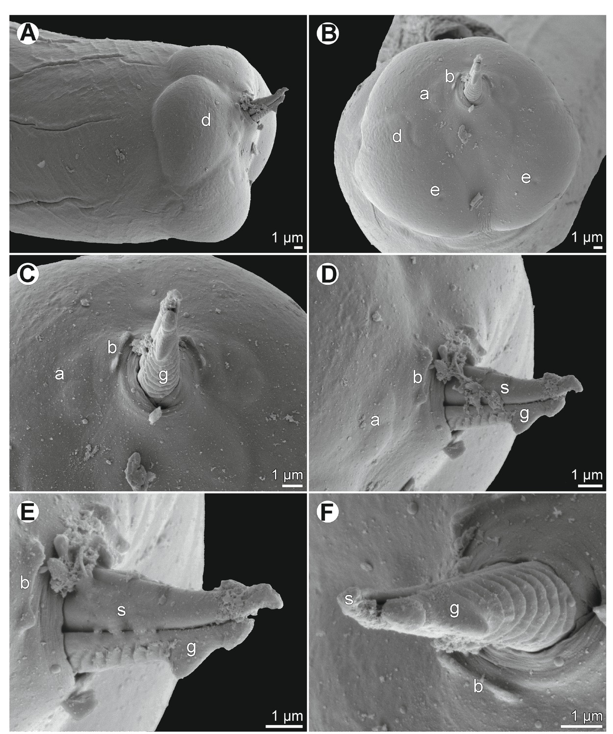
Fig. 6
Philometra consimilis sp. n. from T. crocodilus, scanning electron micrographs of male. A, B – caudal end, lateral and apical views, respectively; C, D – enlarged region of cloaca, apical and sublateral views, respectively; E, F – distal tip of gubernaculum, lateral and subdorsal views, respectively. Abbreviations: a – two flat adanal papillae; b – two elevated adanal papillae; d – caudal mound; e – cuticular depression; g – gubernaculum; s – spicule