
Table 1
Hematological and serum biochemical parameters of a 12-year old mixed breed cat with heartworm infection.
| Parameter | Measure unit | Reference value | Results (interpretation*) |
|---|---|---|---|
| White blood cells (WBC) | K/μL | 5.1 – 16.2 | 25.99 (↑) |
| Hematocrit (HCT) | % | 27.7 – 46.8 | 38.9 |
| Hemoglobin (HGB) | g/dL | 8.1 – 14.2 | 13.1 |
| Neutrophils (NEU) | K/μL | 2.3 – 10.7 | 11.09 (↑) |
| Lymphocytes (LYM) | K/μL | 1.2 – 6.8 | 13.6 (↑) |
| Monocytes (MON) | K/μL | 0.1 – 0.4 | 0.54 (↑) |
| Platels (PLT) | K/μL | 156 – 626 | 216 |
| Blood Uruine Nitrogen (BUN) | mg/dL | 15 – 35 | 27 |
| Glucose (GLU) | mg/dL | 61 – 120 | 162 (↑) |
| Alkaline phosphatase (ALKP) | UI/L | 12 – 65 | 118 (↑) |
| Total protein (TP) | g/dL | 5.4 – 7.9 | 8.7 (↑) |
| Alaninaminotranspherase (ALT) | UI/L | 8 – 53 | 67 (↑) |
| Creatinine (CRE) | mg/dL | 0.5 – 2 | 1.5 |
| Amylase (AMYL) | UI/L | 370 – 1200 | 894 |
| Total bilirubin (TBIL) | μmol/L/dL | 0.15 – 0.5 | 0.3 |
[i] * ↑: increased
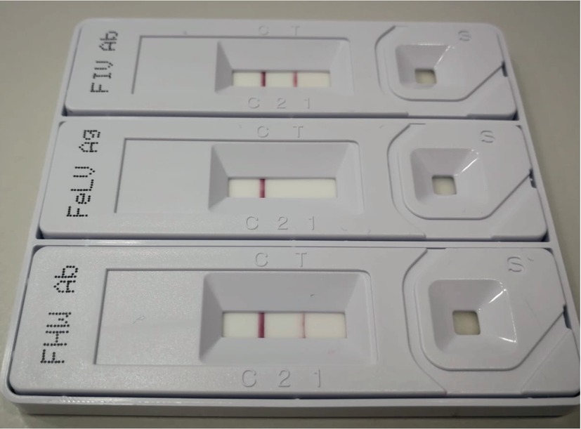
Fig. 1
Serologic (FeliCheck-3) test showing positive reaction for feline immunodeficiency virus (FIV) and feline heartworm (FHW) infection
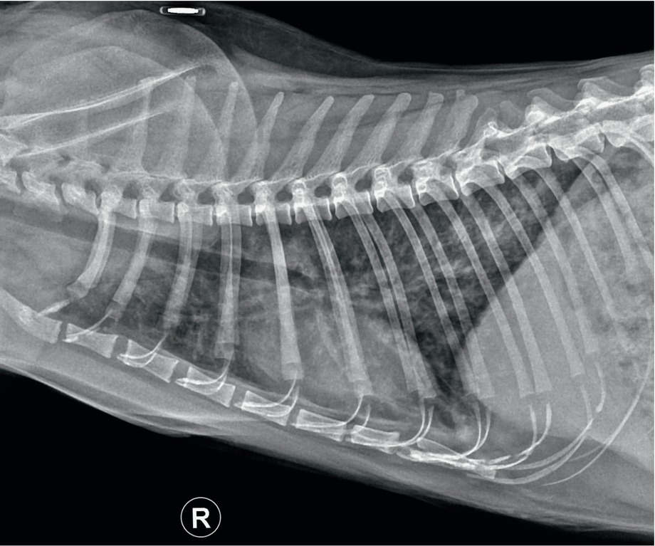
Fig. 2
Thoracic radiograph showing a diffuse bronchointerstitial pattern in a 12-year old cat with heartworm infection (lateral view)
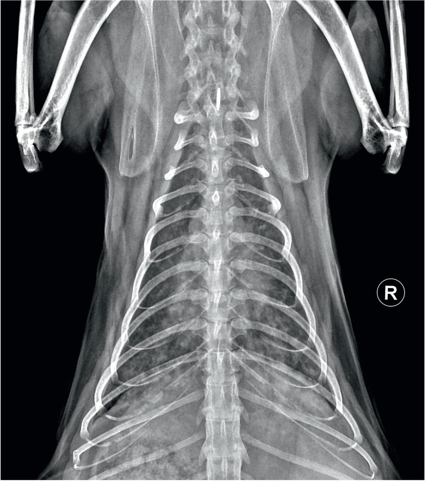
Fig. 3
Thoracic radiograph showing enlarged caudal lobar pulmonary arteries in a 12-year old cat with heartworm infection (dorsoventral view)
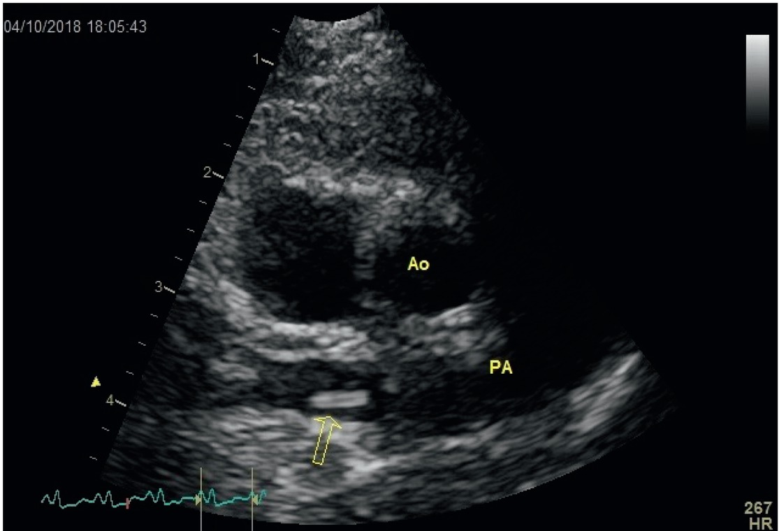
Fig. 4
Echocardiogram showing heartworms, seen as short, segmented, strongly echogenic parallel lines in the right pulmonary artery (arrow) of a 12-year naturally infected cat (Ao: aorta; PA: pulmonary artery) (right parasternal short axis view at the heart’s base)