
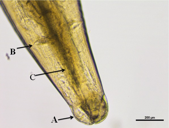
Fig.1
Photomicrograph of anterior end of Toxocara canis showing triradiate lips (A), cervical alae (B) and oesophagus (C) (10×)
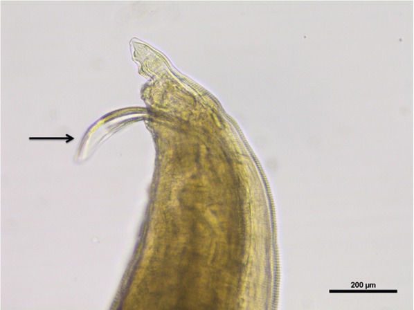
Fig. 2
Photomicrograph of posterior end of male T. canis showing sub equal spicules (arrow) (10×)
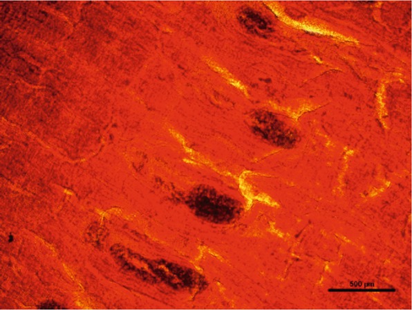
Fig. 3
Photomicrograph of gravid proglottid of Spirometra species exhibiting spiralled uterus (4×)
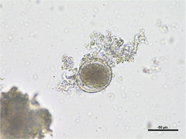
Fig. 4
Photomicrograph of unembryonated Toxocara canis egg (40×)
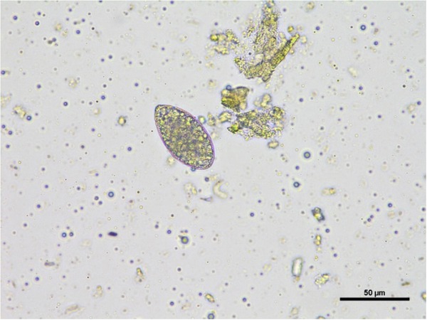
Fig. 5
Photomicrograph of Spirometra species egg with pointed ends (40×)
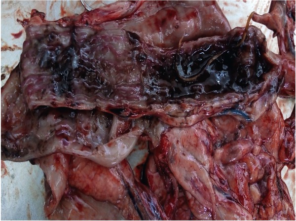
Fig. 6
Intestinal mucosa showing presence of round worms along with catarrhal to haemorrhagic exudate
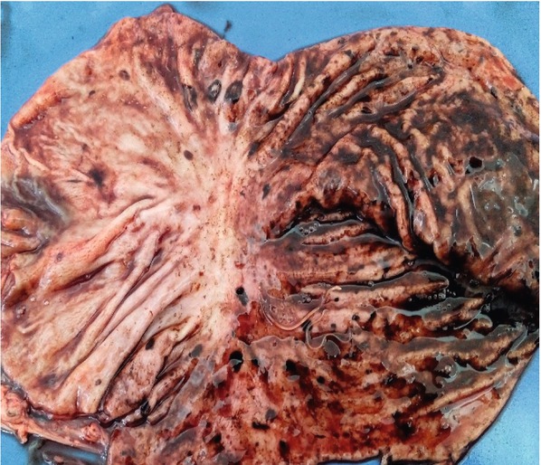
Fig. 7
Thickened gastric mucosa along with multifocal erosive and ulcerative areas
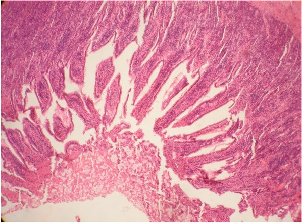
Fig. 8
Photomicrograph showed denuded intestinal epithelium, homogenous pink catarrhal exudates and mononuclear cells admixed with few neutrophils (40×)