
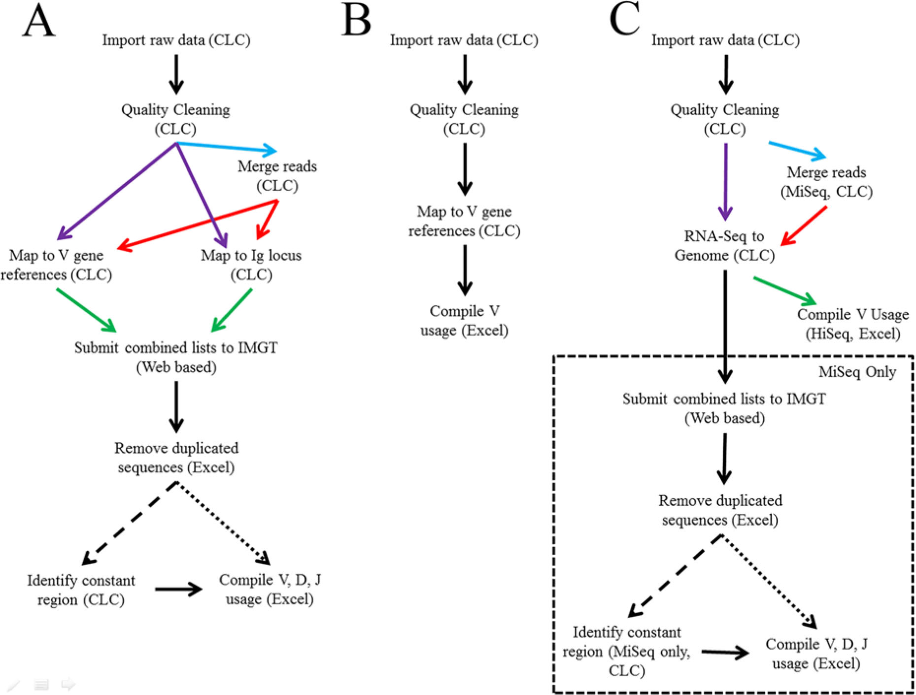
Figure 1
Bioinformatic analysis workflows. (A) Workflow for MiSeq® reference mapping strategy using CLC Genomics Workbench software, the ImMunoGeneTics (IMGT) data base and Excel. (B) Workflow for HiSeq® reference mapping strategy. (C) Workflow for MiSeq® and HiSeq® genome mapping strategy.
Table 1
Sequences used for heavy chain identification. Motifs used to determine the constant region of heavy chain Ig sequences.
| Constant Region | Motif Sequence |
|---|---|
| IgA | GAGTCTGCGAGAAATCCCAC |
| IgD | GTAATGAAAAGGGACCTGAC |
| IgE | TCTATCAGGAACCCTCAGCT |
| IgG1/2b/2c | GCCAAAACAACAGCCCCATC |
| IgG3 | AACAACAGCCCCATCGGTCT |
| IgM | TCAGTCCTTCCCAAATGTCT |
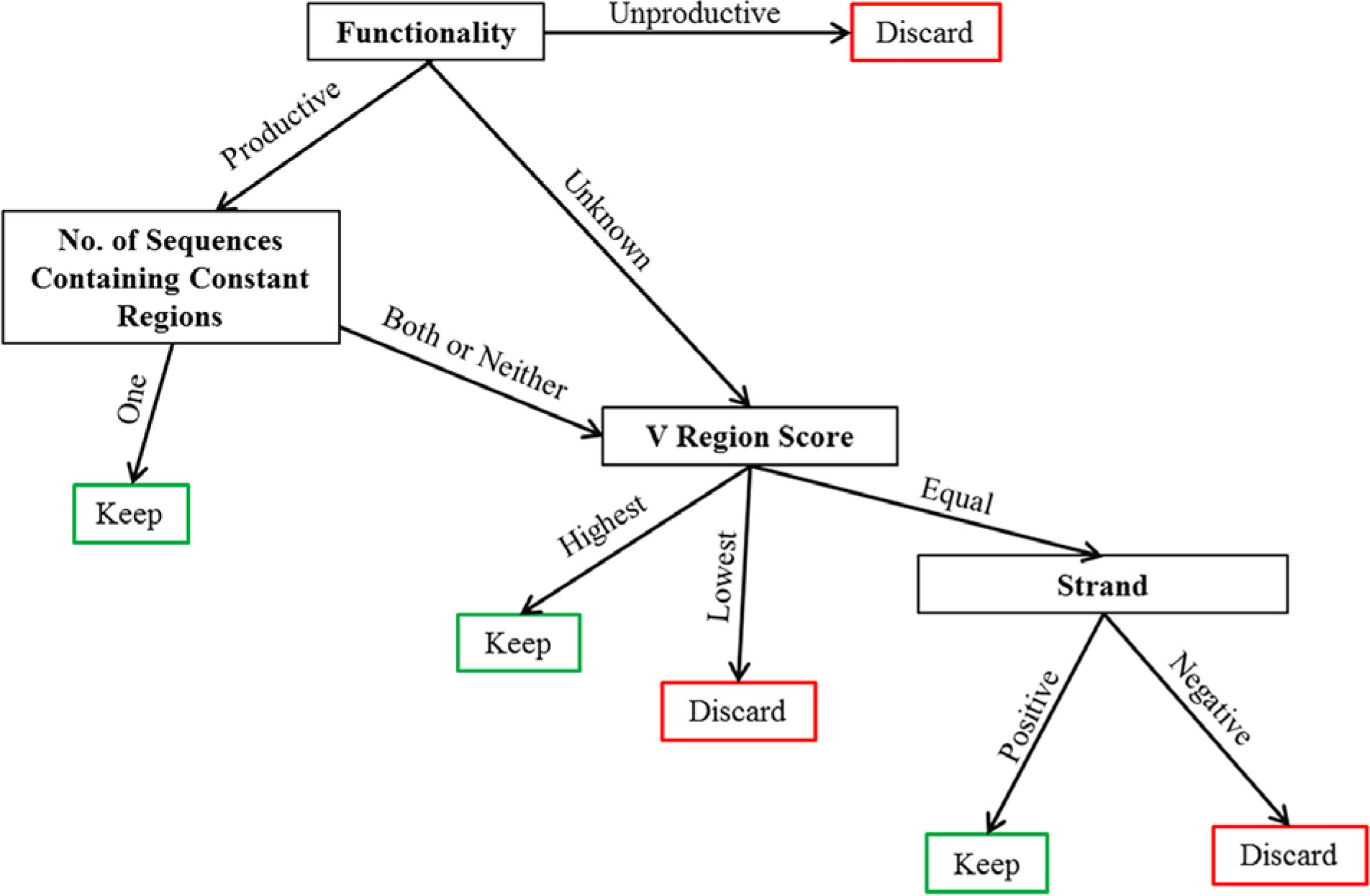
Figure 2
Decision-making matrix to remove duplicate sequence reads after IMGT processing. Mapped sequences that were identified using Illumina sequence identification tags and sequences identified multiple times were removed as outlined.
Table 2
Sequencing and mapping results from the cells, tissue, and size selected treatment groups.
| Cells | Tissue | Size Selected | |
|---|---|---|---|
| Total Reads | 23.9 M | 25.9 M | 21.5 M |
| Post Cleaning | 18.7 M | 20.3 M | 12M |
| IgH Mapped | 278318 | 313194 | 327015 |
| VH Mapped | 12851 | 26559 | 42375 |
| Igκ Mapped | 261037 | 273562 | 264938 |
| Vκ Mapped | 20776 | 35719 | 64540 |
| Heavy Chain Productive | 2036 | 4991 | 8939 |
| Heavy Chain Unknown | 6139 | 11374 | 14047 |
| Light Chain Productive | 3439 | 6799 | 11595 |
| Light Chain Unknown | 6894 | 10462 | 12393 |
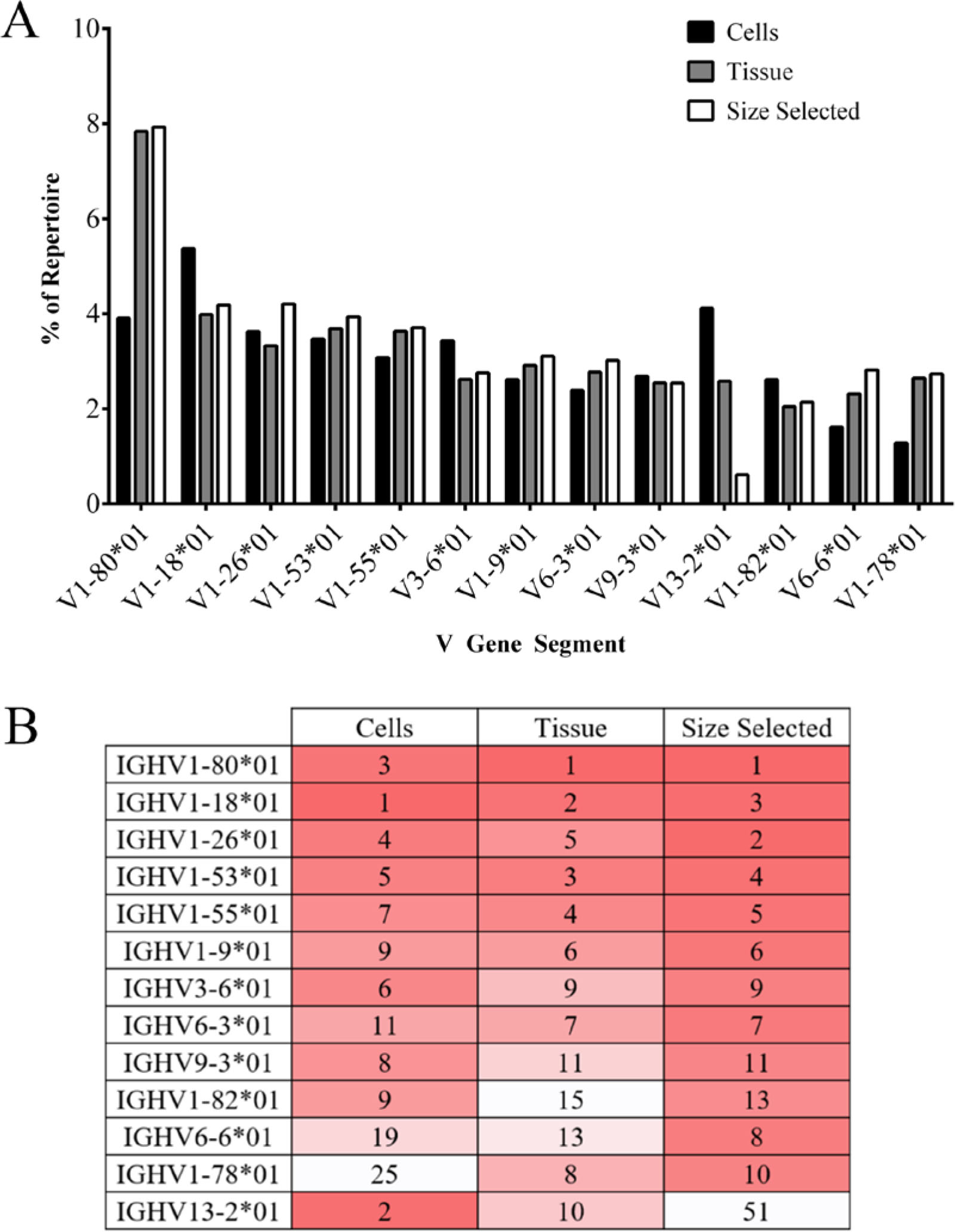
Figure 3
Top ten VH gene segments used among treatment groups. (A) The top ten VH gene segments for each treatment group are presented as a percent of repertoire with corresponding percent of repertoire in other treatment groups listed (because there is not complete overlap among the three treatments groups, 13 VH are presented). (B) Top VH gene segments are listed by rank order (most frequent to least frequent). Dark red indicates higher rank moving to white, of lower rank. VH-gene segments with identical ranks are displayed as ties.
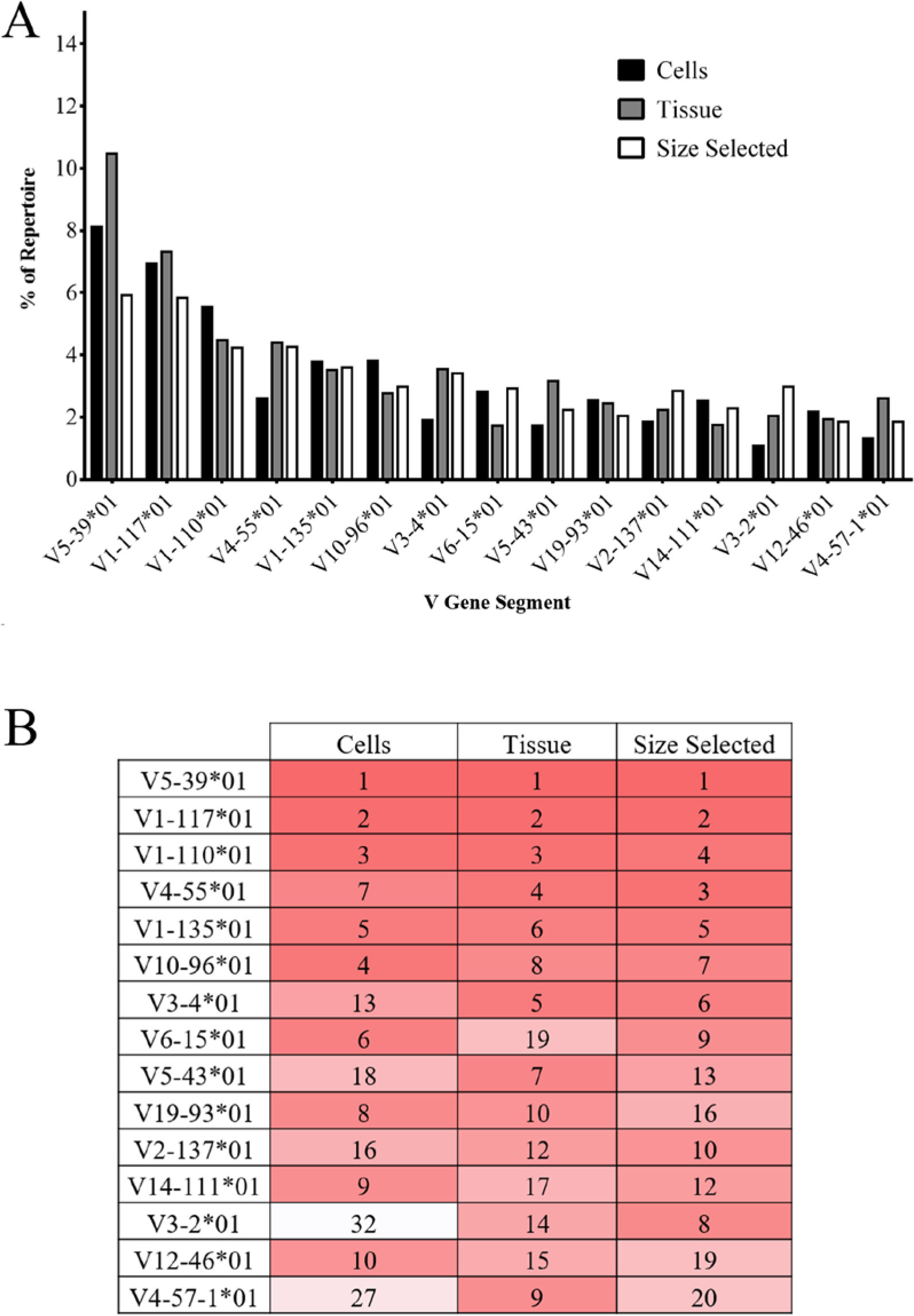
Figure 4
Top ten Vκ used among treatment groups. (A) The top ten Vκ gene segments for each treatment group are presented as a percent of repertoire with corresponding percent of repertoire in other treatment groups listed (because there is not complete overlap among the three treatment groups, 15 Vκ are presented). (B) The top Vκ gene segments are listed by rank order (most frequent to least frequent). Dark red indicates higher rank moving to white, lower rank. VH-gene segments with identical ranks are displayed as ties.
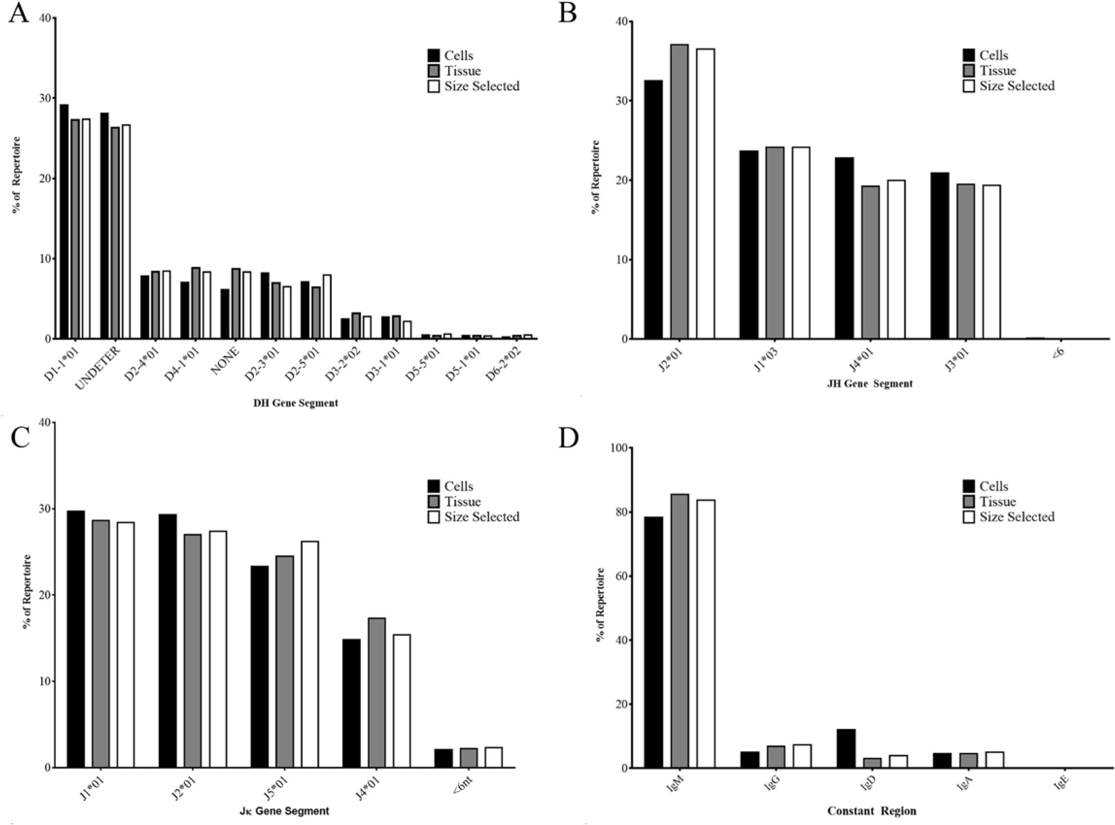
Figure 5
D, J, and heavy chain constant usage among treatment groups. (A) DH gene segment usage by percent of repertoire. (B) JH gene segment usage by percent of repertoire. (C) Jκ gene segment usage by percent of repertoire. (D) Heavy chain constant region usage by percent of repertoire.
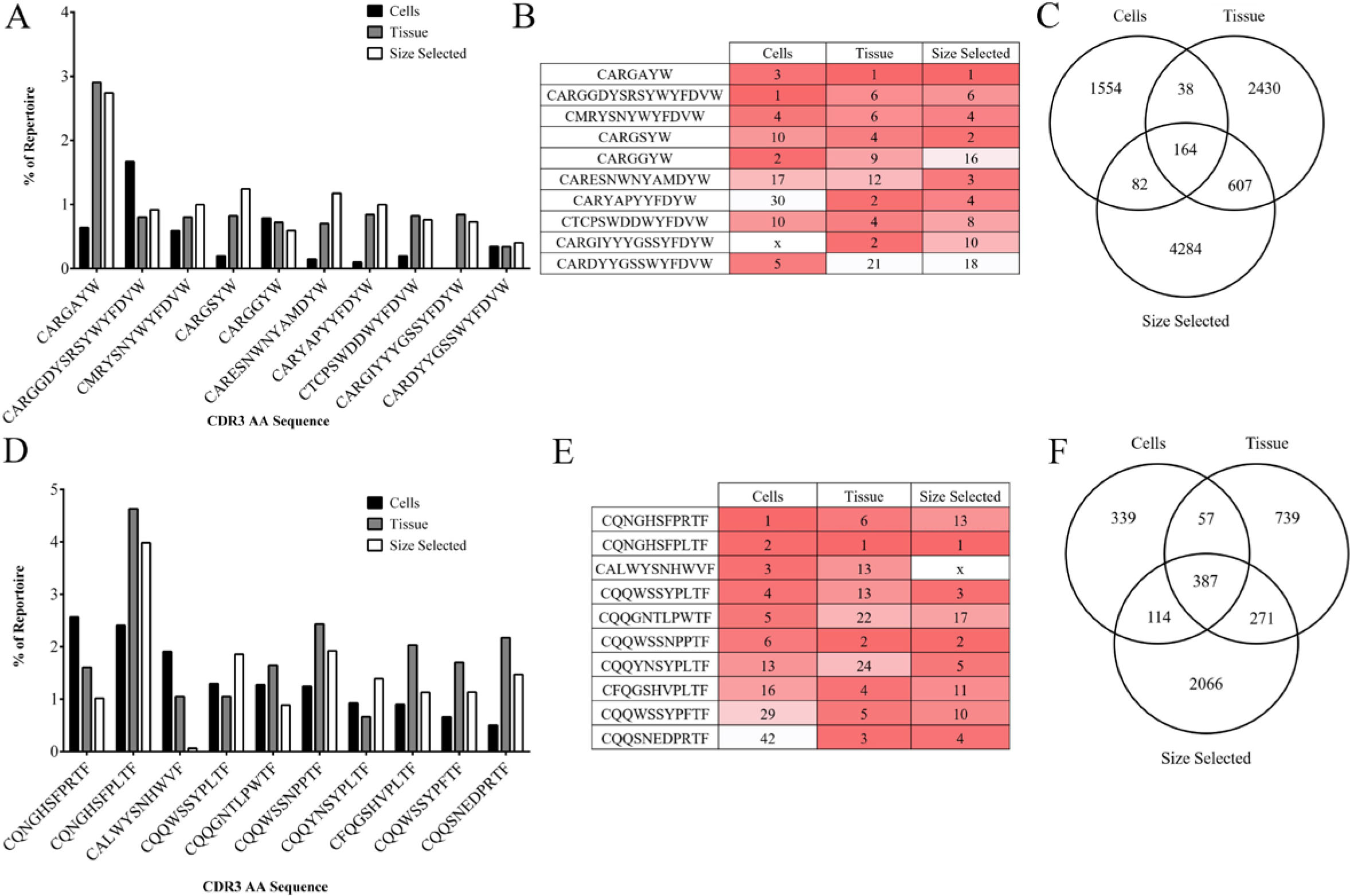
Figure 6
CDR3 AA sequence usage among treatment groups. (A) Usage of the top five most common heavy chain CDR3 AA sequences from each treatment group are presented as percent of repertoire (because there is not complete overlap among the three treatment groups, 10 IgH CDR3s are presented). (B) Most common heavy chain CDR3 AA sequence usage presented by rank. Dark red indicates higher rank moving to white, of lower rank. An x denotes that the AA sequence was not found. (C) Unique heavy chain CDR3 AA sequences identified within and among treatment groups. (D) Usage of the top five most common kappa chain CDR3 AA sequences are presented as percent of repertoire (because there is not complete overlap among the three treatment groups, 10 Igκ CDR3s are presented). (E) Most common heavy chain CDR3 AA sequence usage presented by rank. (F) Unique heavy chain CDR3 AA sequences identified within and among treatment groups.
Table 3
Comparison of mapping techniques in HiSeq® datasets. Mapping techniques were compared by assessing the correlation of Vκ usage between multiple HiSeq® and MiSeq® datasets. HiSeq® datasets included sequencing data from CASIS and NASA ground (G) or flight (F) RR1 mice. The comparison groups are as follows:
| Reference1 | Genome2 | Compared3 | ||
|---|---|---|---|---|
| Cohort | N | R2 (p-value) | R2 (p-value) | R2 (p-value) |
| CASIS G | 3 | 0.030 (.0637) | 0.101 (0.0015) | 0.011 (.027) |
| CASIS F | 3 | 0.001 (.776) | 0.216 (<.0001) | 0.042 (.074) |
| NASA G | 7 | < 0.001 (.854) | 0.379 (<.0001) | 0.013 (.262) |
| NASA F | 7 | 0.004 (.521) | 0.277 (<.0001) | 0.006 (.476) |
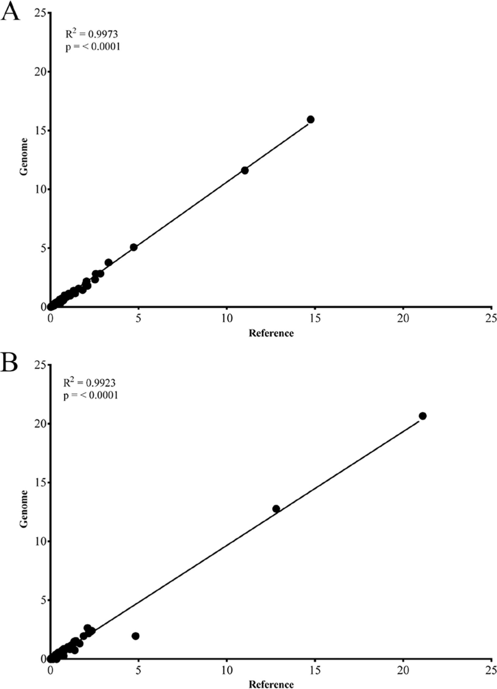
Figure 7
Correlation of V-gene segments between genome and reference mapping. (A) Linear regression of median VH gene segment usage from genome and reference mappings. R2=0.9973, p<0.0001. (B) Linear Regression of median Vκ gene segment usage from genome and reference mapping. R2=0.9923, p<0.0001.